Acta Phys. -Chim. Sin. ›› 2023, Vol. 39 ›› Issue (3): 2210017.doi: 10.3866/PKU.WHXB202210017
• REVIEW • Previous Articles Next Articles
Four-Dimensional Scanning Transmission Electron Microscopy: From Material Microstructures to Physicochemical Properties
Qilong Feng, Chongzhi Zhu, Guan Sheng, Tulai Sun, Yonghe Li, Yihan Zhu( )
)
- Electron Microscopy Center (Analysis and Testing Center), Frontier Interdisciplinary Research Institute, School of Chemical Engineering, Zhejiang University of Technology, Hangzhou 310014, China
-
Received:2022-10-13Accepted:2022-11-29Published:2022-12-05 -
Contact:Yihan Zhu E-mail:yihanzhu@zjut.edu.cn -
About author:Yihan Zhu, Email: yihanzhu@zjut.edu.cn
-
Supported by:the National Key Research and Development Program of China(2022YFE0113800);the National Natural Science Foundation of China(22075250);the National Natural Science Foundation of China(22122505);the National Natural Science Foundation of China(21771161)

Cite this article
Qilong Feng, Chongzhi Zhu, Guan Sheng, Tulai Sun, Yonghe Li, Yihan Zhu. Four-Dimensional Scanning Transmission Electron Microscopy: From Material Microstructures to Physicochemical Properties[J]. Acta Phys. -Chim. Sin. 2023, 39(3), 2210017. doi: 10.3866/PKU.WHXB202210017
share this article
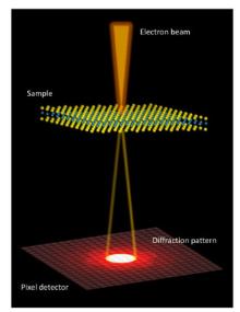
Fig 1
Schematic diagram of the experimental setup for 4D-STEM technology."
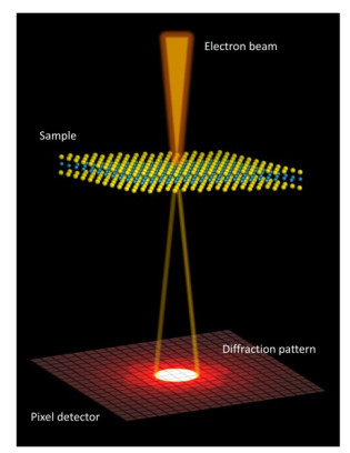
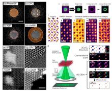
Fig 2
Virtual detector imaging using 4D-STEM technology20, 23, 30. (a–d) Four detector configurations. (a) Bright-field detector. (b) Annular bright field detector. (c) Four-division detector. (d) Low angle annular dark field detector. (e) Reconstructed MoS2 brightfield image. (f) Reconstructed MoS2 annular bright field image. (g) Reconstructed MoS2 differential phase contrast image. (h) Reconstructed MoS2 low-angle annular dark field image. (i) Demonstration of different masked regions for virtual detectors. (j) DyScO3 atomic model schematic. (k) HAADF image generated by hardware high angle annular dark field detector. (l) Reconstructed DyScO3 brightfield image. (m) Reconstructed DyScO3 annular dark field image. (n) Reconstructed DyScO3 annular bright field image. (o) Schematic diagram of 4D-STEM data acquisition (left), images reconstructed from 4D-STEM data, including HAADF image, integrated phase contrast image, differential phase contrast image, and center of mass image. (a–h) Adapted from American Chemical Society publisher. (i–n) Adapted from Springer Nature publisher. (o) Adapted from Springer Nature publisher."
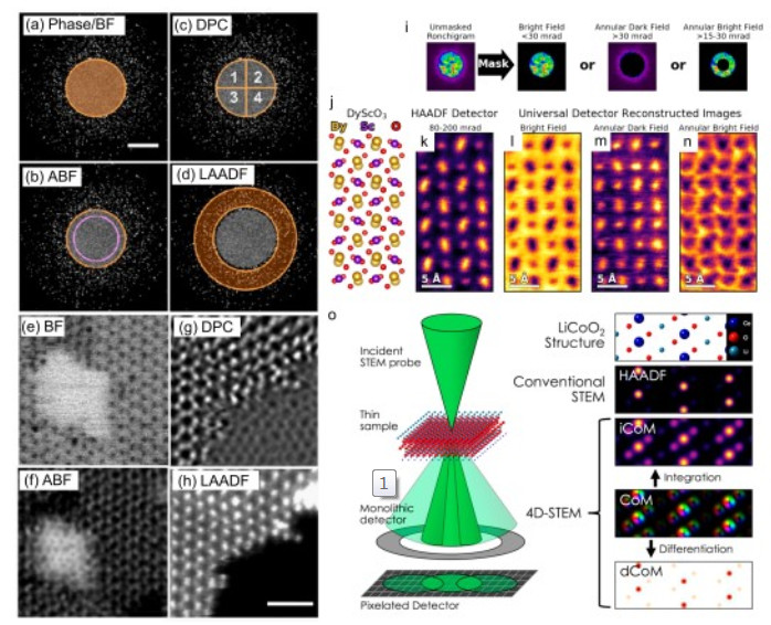
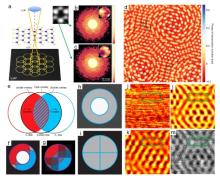
Fig 3
Electron ptychography imaging using 4D-STEM technology39, 41. (a) The basic configuration of the defocus ptychography imaging. (b, c) Converging beam electron diffraction pattern at two locations next to the atomic column, inset showing the intensity distribution inside the bright-field disk. (d) Phase contrast image of bilayer MoS2 reconstructed by electron ptychography imaging technique. (e) Schematic of the overlap of the diffractive disks, red and blue represent opposite phases, so only two overlapping regions are non-zero values. (f) The relative distribution of the diffracted disk on the annular brightfield detector. (g) Relative distribution of diffraction Disk on the four-division detector. (h) Annular bright-field detector configuration. (i) Four-division detector configuration. (j) Annular bright-field image of bilayer graphene. (k) Integral phase contrast image of bilayer graphene. (l) SSB-ptychography of bilayer graphene reconstructs phase contrast images. (m) High-angle annular dark-field image of double-layer graphene. The difference in contrast of the green dotted box is obvious. (a–d) Adapted from Springer Nature publisher. (e–m) Adapted from Elsevier publisher."
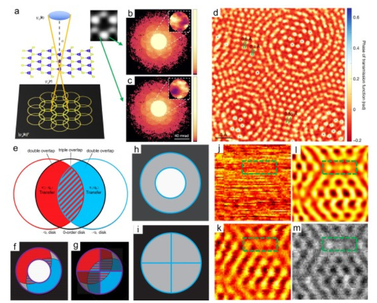
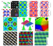
Fig 4
Using 4D-STEM technology to obtain the internal electromagnetic field and valence state distribution of the material54–57. (a) SrTiO3 atomic model. (b) BiFeO3 atomic model. (c) High-angle annular dark field image of SrTiO3. (d) High-angle annular dark-field image of BiFeO3, with inset indicating the offset of Bi atoms. (e) Reconstructed SrTiO3 electric field distribution map. (f) Reconstructed BiFeO3 electric field distribution map. (g) Reconstructed SrTiO3 charge density distribution map. (h) Reconstructed BiFeO3 charge density distribution map. (i) The charge density distribution of SrTiO3 calculated by density functional theory. (j) The charge density distribution of BiFeO3 calculated by density functional theory. (k) Valence electron map of TiO2 aspheric surface. (l) STEM schematic of an electron beam passing through a uniformly magnetized thin film. (m) Color wheel diagram of the magnetic field, showing the direction and magnitude of the magnetic field in the plane. (n) The ADF image of α-Fe2O3 observed along the [${\rm{\bar 1}}$${\rm{\bar 1}}$20] direction. (o) Projected magnetic field vector colour map of α-Fe2O3. The inset color wheel indicates how colour and shade denote the magnetic field orientation and strength in the vector colour map. (p) The magnetic phase-shift image of α-Fe2O3. (a–j) Adapted from Springer Nature publisher. (k) Adapted from Elsevier publisher. (l–m) Adapted from Elsevier publisher. (n–p) Adapted from Springer Nature publisher."
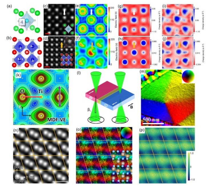
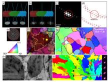
Fig 5
Determination of crystal orientation of materials by 4D-STEM technology9, 70–73. (a) The Kikuchi line was used to determine the crystal orientation. (b) The crystal orientation was determined by the Bragg diffraction point. (c) Crystal diffraction peaks of single crystal silicon under the < 110 > axis. (d) Matching bragg diffraction peaks based on cross-correlation template matching. (e) The Kikuchi diffraction pattern at the yellow dot in (f). (f) Reconstruction of crystal orientation maps of tungsten films using the Kikuchi line fitting method. (g) Reconstruction of crystal orientation map of single-crystal silicon thin films by the method of calibrating Bragg diffraction points. (h) Annular dark field image of the thin film. (i) The crystal orientation distribution map corresponding to the region in Fig.(h). The right side is the diffraction pattern in the corresponding marked range. (a, b) Adapted from Cambridge University Press publisher. (c, d) Adapted from Elsevier publisher. (e, f) Adapted from Cambridge University Press publisher. (g) Adapted from Cambridge University Press publisher. (h, i) Adapted from Springer Nature publisher."
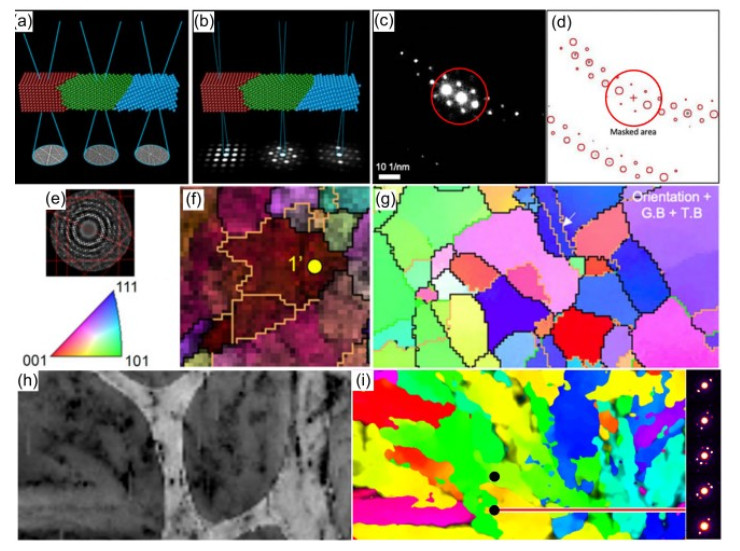
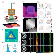
Fig 6
Determination of internal stress distribution of materials by 4D-STEM technology77, 79–81. (a) Experimental configuration of the nanobeam electron diffraction. (b) Image of Rh@Pt nanocube after correction the scan distortion and the corrected scan positions are marked as blue dots (c) Mapping of normal and shear strains of Rh@Pt nanocubes, the lower left corner of each strain diagram represents the geometric interpretation of strain formation. (d) Image of Pd@Pt core-shell nanoparticles. (e) Mapping of normal strain and shear strain of Pd@Pt core-shell nanoparticles. (f) Uniaxial strain map. (g) Strain histogram from (f). The inset shows the histogram from a flat region (gray box in panel f) indicating a precision better than ∼0.18%. (h) The average diffraction pattern and a virtual dark-field image are shown along with the color-coded strain maps. Three time resolved maps are shown. The sample is deformed under continuous loading before showing catastrophic fractur. (a–c) Adapted from American Chemical Society publisher. (d, e) Adapted from Elsevier publisher. (f, g) Adapted from American Chemical Society publisher. (h) Adapted from AIP Publishing publisher."
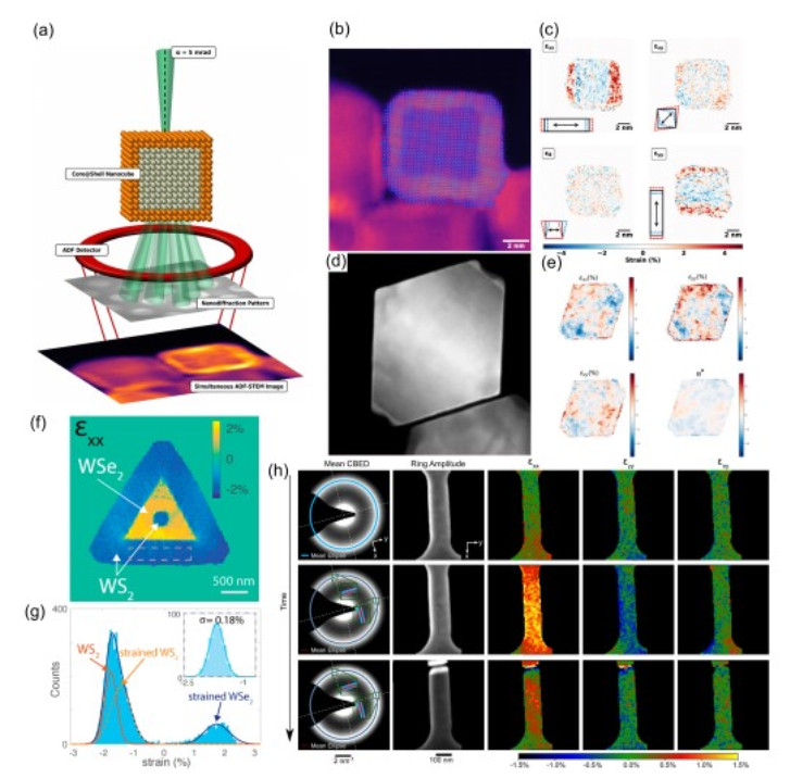
Fig 7
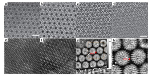
Application of 4D-STEM technology in low dose field. Using the same set of 4D-STEM data collected on the ZSM-5 sample to reconstruct40, 46, 102. (a) The annular dark field image, (b) integrate phase contrast image, (c) single-side band electron ptychography image of ZSM-5, (d) single-side band electron ptychography image of ZSM-5 with a cumulative electron dose of 200 e∙Å?2. Using the same set of 4D-STEM data collected on the bilayer MoSe2/WS2 sample to reconstruct (e) the annular dark field image, (f) iterative electron ptychography image. (g) Iterative electron ptychography image of viral protein with cumulative electron dose of 22.8 e∙Å?2. (h) Enlarged image within the orange boxed area in Fig. 7g. (a–d) Adapted from AIP Publishing publisher. (e, f) Adapted from Springer Nature publisher. (g, h) Adapted from Springer Nature publisher."
| 1 |
Kisielowski C. ; Freitag B. ; Bischoff M. ; van Lin H. ; Lazar S. ; Knippels G. ; Tiemeijer P. ; van der Stam M. ; von Harrach S. ; Stekelenburg M. ; et al Microsc. Microanal. 2008, 14 (5), 469.
doi: 10.1017/S1431927608080902 |
| 2 |
Yankovich A. B. ; Berkels B. ; Dahmen W. ; Binev P. ; Sanchez S. I. ; Bradley S. A. ; Li A. ; Szlufarska I. ; Voyles P. M. Nat. Commun. 2014, 5, 4155.
doi: 10.1038/ncomms5155 |
| 3 | Lazic, I.; Bosch, E. G. T. Advances in Imaging and Electron Physics. Hawkes, P. W., Ed. Elsevier: Toulouse, France, 2017; Vol. 199, pp. 303–309. |
| 4 |
Lazic I. ; Bosch E. G. T. ; Lazar S. Ultramicroscopy 2016, 160, 265.
doi: 10.1016/j.ultramic.2015.10.011 |
| 5 |
Yucelen E. ; Lazic I. ; Bosch E. G. T. Sci. Rep. 2018, 8 (1), 2676.
doi: 10.1038/s41598-018-20377-2 |
| 6 |
Seifer S. ; Houben L. ; Elbaum M. Microsc. Microanal. 2021, 27, 1476.
doi: 10.1017/S1431927621012861 |
| 7 |
Nord M. ; Webster R. W. H. ; Paton K. A. ; McVitie S. ; McGrouther D. ; MacLaren I. ; Paterson G. W. Microsc. Microanal. 2020, 26 (4), 653.
doi: 10.1017/S1431927620001713 |
| 8 |
Tate M. W. ; Purohit P. ; Chamberlain D. ; Nguyen K. X. ; Hovden R. ; Chang C. S. ; Deb P. ; Turgut E. ; Heron J. T. ; Schlom D. G. ; et al Microsc. Microanal. 2016, 22 (1), 237.
doi: 10.1017/S1431927615015664 |
| 9 |
Ophus C. Microsc. Microanal. 2019, 25 (3), 563.
doi: 10.1017/S1431927619000497 |
| 10 |
Levin B. D. ; Zhang C. ; Bammes B. ; Voyles P. M. ; Bilhorn R. B. Microsc. Anal. 2020, 34, 20.
doi: 10.1017/S1431927621003809 |
| 11 |
Nellist P. D. ; McCallum B. C. ; Rodenburg J. M. Nature 1995, 374 (6523), 630.
doi: 10.1038/374630a0 |
| 12 |
Savitzky B. H. ; Zeltmann S. E. ; Hughes L. A. ; Brown H. G. ; Zhao S. ; Pelz P. M. ; Pekin T. C. ; Barnard E. S. ; Donohue J. ; DaCosta L. R. ; et al Microsc. Microanal. 2021, 27 (4), 712.
doi: 10.1017/S1431927621000477 |
| 13 |
Cautaerts N. ; Crout P. ; Anes H. W. ; Prestat E. ; Jeong J. ; Dehm G. ; Liebscher C. H. Ultramicroscopy 2022, 237, 113517.
doi: 10.1016/j.ultramic.2022.113517 |
| 14 |
Zaluzec N. J. Microsc. Microanal. 2002, 8 (S02), 376.
doi: 10.1017/S143192760210064X |
| 15 |
Ozdol V. B. ; Gammer C. ; Jin X. G. ; Ercius P. ; Ophus C. ; Ciston J. ; Minor A. M. Appl. Phys. Lett. 2015, 106 (25), 253107.
doi: 10.1063/1.4922994 |
| 16 |
Liu A. C. ; Neish M. J. ; Stokol G. ; Buckley G. A. ; Smillie L. A. ; de Jonge M. D. ; Ott R. T. ; Kramer M. J. ; Bourgeois L. Phys. Rev. Lett. 2013, 110 (20), 205505.
doi: 10.1103/PhysRevLett.110.205505 |
| 17 |
Kimoto K. ; Ishizuka K. Ultramicroscopy 2011, 111 (8), 1111.
doi: 10.1016/j.ultramic.2011.01.029 |
| 18 |
Jarausch K. ; Thomas P. ; Leonard D. N. ; Twesten R. ; Booth C. R. Ultramicroscopy 2009, 109 (4), 326.
doi: 10.1016/j.ultramic.2008.12.012 |
| 19 |
Yedra L. ; Eljarrat A. ; Arenal R. ; Pellicer E. ; Cabo M. ; Lopez-Ortega A. ; Estrader M. ; Sort J. ; Baro M. D. ; Estrade S. ; et al Ultramicroscopy 2012, 122, 12.
doi: 10.1016/j.ultramic.2012.07.020 |
| 20 |
Hachtel J. A. ; Idrobo J. C. ; Chi M. Adv. Struct. Chem. Imag. 2018, 4 (1), 10.
doi: 10.1186/s40679-018-0059-4 |
| 21 |
Bosch E. G. ; Lazic I. Ultramicroscopy 2015, 156, 59.
doi: 10.1016/j.ultramic.2015.02.004 |
| 22 |
Lupini A. R. ; Chi M. ; Kalinin S. V. ; Borisevich A. Y. ; Carlos Idrobo J. ; Jesse S. Microsc. Microanal. 2015, 21 (S3), 1219.
doi: 10.1017/s1431927615006881 |
| 23 |
Wen Y. ; Ophus C. ; Allen C. S. ; Fang S. ; Chen J. ; Kaxiras E. ; Kirkland A. I. ; Warner J. H. Nano Lett. 2019, 19 (9), 6482.
doi: 10.1021/acs.nanolett.9b02717 |
| 24 |
Nellist P. D. ; Chisholm M. F. ; Dellby N. ; Krivanek O. L. ; Murfitt M. F. ; Szilagyi Z. S. ; Lupini A. R. ; Borisevich A. ; Sides W. H. ; Pennycook S. J. Science 2004, 305 (5691), 1741.
doi: 10.1126/science.1100965 |
| 25 |
Ishikawa R. ; Okunishi E. ; Sawada H. ; Kondo Y. ; Hosokawa F. ; Abe E. Nat. Mater. 2011, 10 (4), 278.
doi: 10.1038/nmat2957 |
| 26 |
Ohtsuka M. ; Yamazaki T. ; Kotaka Y. ; Hashimoto I. ; Watanabe K. Ultramicroscopy 2012, 120, 48.
doi: 10.1016/j.ultramic.2012.06.006 |
| 27 |
Findlay S. D. ; Kohno Y. ; Cardamone L. A. ; Ikuhara Y. ; Shibata N. Ultramicroscopy 2014, 136, 31.
doi: 10.1016/j.ultramic.2013.07.019 |
| 28 |
Ahmed S. ; Bianchini M. ; Pokle A. ; Munde M. S. ; Hartmann P. ; Brezesinski T. ; Beyer A. ; Janek J. ; Volz K. Adv. Energy Mater. 2020, 10 (25), 2001026.
doi: 10.1002/aenm.202001026 |
| 29 |
de Graaf S. ; Momand J. ; Mitterbauer C. ; Lazar S. ; Kooi B. J. Sci. Adv. 2020, 6 (5), eaay4312.
doi: 10.1126/sciadv.aay4312 |
| 30 |
Xiong H. ; Liu Z. ; Chen X. ; Wang H. ; Qian W. ; Zhang C. ; Zheng A. ; Wei F. Science 2022, 376 (6592), 491.
doi: 10.1126/science.abn7667 |
| 31 |
Muller K. ; Krause F. F. ; Beche A. ; Schowalter M. ; Galioit V. ; Loffler S. ; Verbeeck J. ; Zweck J. ; Schattschneider P. ; Rosenauer A. Nat. Commun. 2014, 5, 5653.
doi: 10.1038/ncomms6653 |
| 32 |
MacLaren I. ; Wang L. ; McGrouther D. ; Craven A. J. ; McVitie S. ; Schierholz R. ; Kovacs A. ; Barthel J. ; Dunin-Borkowski R. E. Ultramicroscopy 2015, 154, 57.
doi: 10.1016/j.ultramic.2015.03.016 |
| 33 |
Shibata N. ; Findlay S. D. ; Kohno Y. ; Sawada H. ; Kondo Y. ; Ikuhara Y. Nat. Phys. 2012, 8 (8), 611.
doi: 10.1038/nphys2337 |
| 34 |
Zachman M. J. ; Yang Z. ; Du Y. ; Chi M. ACS Nano 2022, 16 (1), 1358.
doi: 10.1021/acsnano.1c09374 |
| 35 |
Ooe K. ; Seki T. ; Ikuhara Y. ; Shibata N. Ultramicroscopy 2019, 202, 148.
doi: 10.1016/j.ultramic.2019.04.011 |
| 36 |
Yu C.-P. ; Friedrich T. ; Jannis D. ; Van Aert S. ; Verbeeck J. Microsc. Microanal. 2022, 28 (5), 1526.
doi: 10.1017/S1431927622000617 |
| 37 |
Strauch A. ; Weber D. ; Clausen A. ; Lesnichaia A. ; Bangun A. ; März B. ; Lyu F. J. ; Chen Q. ; Rosenauer A. ; Dunin-Borkowski R. ; et al Microsc. Microanal. 2021, 27 (5), 1078.
doi: 10.1017/s1431927621012423 |
| 38 |
Yang H. ; MacLaren I. ; Jones L. ; Martinez G. T. ; Simson M. ; Huth M. ; Ryll H. ; Soltau H. ; Sagawa R. ; Kondo Y. ; et al Ultramicroscopy 2017, 180, 173.
doi: 10.1016/j.ultramic.2017.02.006 |
| 39 |
Pennycook T. J. ; Lupini A. R. ; Yang H. ; Murfitt M. F. ; Jones L. ; Nellist P. D. Ultramicroscopy 2015, 151, 160.
doi: 10.1016/j.ultramic.2014.09.013 |
| 40 |
Chen Z. ; Odstrcil M. ; Jiang Y. ; Han Y. ; Chiu M. H. ; Li L. J. ; Muller D. A. Nat. Commun. 2020, 11 (1), 2994.
doi: 10.1038/s41467-020-16688-6 |
| 41 |
Jiang Y. ; Chen Z. ; Han Y. ; Deb P. ; Gao H. ; Xie S. ; Purohit P. ; Tate M. W. ; Park J. ; Gruner S. M. ; et al Nature 2018, 559 (7714), 343.
doi: 10.1038/s41586-018-0298-5 |
| 42 |
Yang H. ; Rutte R. N. ; Jones L. ; Simson M. ; Sagawa R. ; Ryll H. ; Huth M. ; Pennycook T. J. ; Green M. L. ; Soltau H. ; et al Nat. Commun. 2016, 7, 12532.
doi: 10.1038/ncomms12532 |
| 43 |
Song J. ; Allen C. S. ; Gao S. ; Huang C. ; Sawada H. ; Pan X. ; Warner J. ; Wang P. ; Kirkland A. I. Sci. Rep. 2019, 9 (1), 3919.
doi: 10.1038/s41598-019-40413-z |
| 44 |
Maiden A. ; Johnson D. ; Li P. Optica 2017, 4 (7), 736.
doi: 10.1364/optica.4.000736 |
| 45 |
Rodenburg J. M. ; Hurst A. C. ; Cullis A. G. Ultramicroscopy 2007, 107 (2–3), 227.
doi: 10.1016/j.ultramic.2006.07.007 |
| 46 |
Zhou L. ; Song J. ; Kim J. S. ; Pei X. ; Huang C. ; Boyce M. ; Mendonca L. ; Clare D. ; Siebert A. ; Allen C. S. ; et al Nat. Commun. 2020, 11 (1), 2773.
doi: 10.1038/s41467-020-16391-6 |
| 47 |
Chen Z. ; Jiang Y. ; Shao Y. T. ; Holtz M. E. ; Odstrcil M. ; Guizar-Sicairos M. ; Hanke I. ; Ganschow S. ; Schlom D. G. ; Muller D. A. Science 2021, 372 (6544), 826.
doi: 10.1126/science.abg2533 |
| 48 |
Maiden A. M. ; Humphry M. J. ; Rodenburg J. M. J. Opt. Soc. Am. A 2012, 29 (8), 1606.
doi: 10.1364/JOSAA.29.001606 |
| 49 |
Hue F. ; Rodenburg J. M. ; Maiden A. M. ; Midgley P. A. Ultramicroscopy 2011, 111 (8), 1117.
doi: 10.1016/j.ultramic.2011.02.005 |
| 50 |
Maiden A. M. ; Rodenburg J. M. Ultramicroscopy 2009, 109 (10), 1256.
doi: 10.1016/j.ultramic.2009.05.012 |
| 51 |
Faulkner H. M. ; Rodenburg J. M. Phys. Rev. Lett. 2004, 93 (2), 023903.
doi: 10.1103/PhysRevLett.93.023903 |
| 52 |
Rodenburg J. M. ; McCallum B. C. ; Nellist P. D. Ultramicroscopy 1993, 48 (3), 304.
doi: 10.1016/0304-3991(93)90105-7 |
| 53 |
Pennycook T. J. ; Martinez G. T. ; Nellist P. D. ; Meyer J. C. Ultramicroscopy 2019, 196, 131.
doi: 10.1016/j.ultramic.2018.10.005 |
| 54 |
Gao W. ; Addiego C. ; Wang H. ; Yan X. ; Hou Y. ; Ji D. ; Heikes C. ; Zhang Y. ; Li L. ; Huyan H. ; et al Nature 2019, 575 (7783), 480.
doi: 10.1038/s41586-019-1649-6 |
| 55 |
Wu L. ; Meng Q. ; Zhu Y. Ultramicroscopy 2020, 219, 113095.
doi: 10.1016/j.ultramic.2020.113095 |
| 56 |
Krajnak M. ; McGrouther D. ; Maneuski D. ; Shea V. O. ; McVitie S. Ultramicroscopy 2016, 165, 42.
doi: 10.1016/j.ultramic.2016.03.006 |
| 57 |
Kohno Y. ; Seki T. ; Findlay S. D. ; Ikuhara Y. ; Shibata N. Nature 2022, 602 (7896), 234.
doi: 10.1038/s41586-021-04254-z |
| 58 |
Yang H. ; Pennycook T. J. ; Nellist P. D. Ultramicroscopy 2015, 151, 232.
doi: 10.1016/j.ultramic.2014.10.013 |
| 59 |
Li Z. ; Biskupek J. ; Kaiser U. ; Rose H. Microsc. Microanal. 2022, 28 (3), 611.
doi: 10.1017/S1431927622000289 |
| 60 |
Fang S. ; Wen Y. ; Allen C. S. ; Ophus C. ; Han G. G. D. ; Kirkland A. I. ; Kaxiras E. ; Warner J. H. Nat. Commun. 2019, 10 (1), 1127.
doi: 10.1038/s41467-019-08904-9 |
| 61 |
Muller-Caspary K. ; Grieb T. ; Mussener J. ; Gauquelin N. ; Hille P. ; Schormann J. ; Verbeeck J. ; Van Aert S. ; Eickhoff M. ; Rosenauer A. Phys. Rev. Lett. 2019, 122 (10), 106102.
doi: 10.1103/PhysRevLett.122.106102 |
| 62 |
Yadav A. K. ; Nguyen K. X. ; Hong Z. ; Garcia-Fernandez P. ; Aguado-Puente P. ; Nelson C. T. ; Das S. ; Prasad B. ; Kwon D. ; Cheema S. ; et al Nature 2019, 565 (7740), 468.
doi: 10.1038/s41586-018-0855-y |
| 63 |
Grieb T. ; Krause F. F. ; Muller-Caspary K. ; Ritz R. ; Simson M. ; Schormann J. ; Mahr C. ; Mussener J. ; Schowalter M. ; Soltau H. ; et al Ultramicroscopy 2021, 228, 113321.
doi: 10.1016/j.ultramic.2021.113321 |
| 64 |
Addiego C. ; Gao W. ; Pan X. Ultramicroscopy 2020, 208, 112850.
doi: 10.1016/j.ultramic.2019.112850 |
| 65 |
Campanini M. ; Nasi L. ; Albertini F. ; Erni R. Appl. Phys. Lett. 2020, 117 (15), 154102.
doi: 10.1063/5.0026121 |
| 66 |
Shibata N. ; Findlay S. D. ; Matsumoto T. ; Kohno Y. ; Seki T. ; Sanchez-Santolino G. ; Ikuhara Y. Acc. Chem. Res. 2017, 50 (7), 1502.
doi: 10.1021/acs.accounts.7b00123 |
| 67 |
Wang B. ; Bagues N. ; Liu T. ; Kawakami R. K. ; McComb D. W. Ultramicroscopy 2022, 232, 113395.
doi: 10.1016/j.ultramic.2021.113395 |
| 68 |
Caplins B. W. ; Holm J. D. ; White R. M. ; Keller R. R. Ultramicroscopy 2020, 219, 113137.
doi: 10.1016/j.ultramic.2020.113137 |
| 69 |
Donohue J. ; Zeltmann S. E. ; Bustillo K. C. ; Savitzky B. ; Jones M. A. ; Meyers G. F. ; Ophus C. ; Minor A. M. iScience 2022, 25 (3), 103882.
doi: 10.1016/j.isci.2022.103882 |
| 70 |
Fundenberger J. J. ; Morawiec A. ; Bouzy E. ; Lecomte J. S. Ultramicroscopy 2003, 96 (2), 127.
doi: 10.1016/s0304-3991(02)00435-7 |
| 71 |
Jeong J. ; Jang W. S. ; Kim K. H. ; Kostka A. ; Gu G. ; Kim Y. M. ; Oh S. H. Microsc. Microanal. 2021, 27 (2), 237.
doi: 10.1017/S1431927621000027 |
| 72 |
Jeong J. ; Cautaerts N. ; Dehm G. ; Liebscher C. H. Microsc. Microanal. 2021, 27 (5), 1102.
doi: 10.1017/s1431927621012538 |
| 73 |
Panova O. ; Ophus C. ; Takacs C. J. ; Bustillo K. C. ; Balhorn L. ; Salleo A. ; Balsara N. ; Minor A. M. Nat. Mater. 2019, 18 (8), 860.
doi: 10.1038/s41563-019-0387-3 |
| 74 |
Bustillo K. C. ; Zeltmann S. E. ; Chen M. ; Donohue J. ; Ciston J. ; Ophus C. ; Minor A. M. Acc. Chem. Res. 2021, 54 (11), 2543.
doi: 10.1021/acs.accounts.1c00073 |
| 75 |
Hÿtch M. J. ; Minor A. M. MRS Bull. 2014, 39 (2), 138.
doi: 10.1557/mrs.2014.4 |
| 76 |
Allen F. I. ; Pekin T. C. ; Persaud A. ; Rozeveld S. J. ; Meyers G. F. ; Ciston J. ; Ophus C. ; Minor A. M. Microsc. Microanal. 2021, 27 (4), 794.
doi: 10.1017/S1431927621011946 |
| 77 |
Han Y. ; Nguyen K. ; Cao M. ; Cueva P. ; Xie S. ; Tate M. W. ; Purohit P. ; Gruner S. M. ; Park J. ; Muller D. A. Nano Lett. 2018, 18 (6), 3746.
doi: 10.1021/acs.nanolett.8b00952 |
| 78 |
Mahr C. ; Muller-Caspary K. ; Grieb T. ; Krause F. F. ; Schowalter M. ; Rosenauer A. Ultramicroscopy 2021, 221, 113196.
doi: 10.1016/j.ultramic.2020.113196 |
| 79 |
Mukherjee D. ; Gamler J. T. L. ; Skrabalak S. E. ; Unocic R. R. ACS Catal. 2020, 10 (10), 5529.
doi: 10.1021/acscatal.0c00224 |
| 80 |
Wang S. ; Eldred T. B. ; Smith J. G. ; Gao W. Ultramicroscopy 2022, 236, 113513.
doi: 10.1016/j.ultramic.2022.113513 |
| 81 |
Gammer C. ; Ophus C. ; Pekin T. C. ; Eckert J. ; Minor A. M. Appl. Phys. Lett. 2018, 112 (17), 1905.
doi: 10.1063/1.5025686 |
| 82 |
Kazmierczak N. P. ; Van Winkle M. ; Ophus C. ; Bustillo K. C. ; Carr S. ; Brown H. G. ; Ciston J. ; Taniguchi T. ; Watanabe K. ; Bediako D. K. Nat. Mater. 2021, 20 (7), 956.
doi: 10.1038/s41563-021-00973-w |
| 83 |
Lebeau J. M. ; Findlay S. D. ; Allen L. J. ; Stemmer S. Ultramicroscopy 2010, 110 (2), 118.
doi: 10.1016/j.ultramic.2009.10.001 |
| 84 |
Ophus C. ; Ercius P. ; Huijben M. ; Ciston J. Appl. Phys. Lett. 2017, 110 (6), 3102.
doi: 10.1063/1.4975932 |
| 85 |
Zhang C. ; Feng J. ; DaCosta L. R. ; Voyles P. M. Ultramicroscopy 2019, 210, 112921.
doi: 10.1016/j.ultramic.2019.112921 |
| 86 |
Chen Q. ; Dwyer C. ; Sheng G. ; Zhu C. ; Li X. ; Zheng C. ; Zhu Y. Adv. Mater. 2020, 32 (16), e1907619.
doi: 10.1002/adma.201907619 |
| 87 |
Bunck D. N. ; Dichtel W. R. J. Am. Chem. Soc. 2013, 135 (40), 14952.
doi: 10.1021/ja408243n |
| 88 |
Zhu Y. ; Ciston J. ; Zheng B. ; Miao X. ; Czarnik C. ; Pan Y. ; Sougrat R. ; Lai Z. ; Hsiung C.-E. ; Yao K. Nat. Mater. 2017, 16 (5), 532.
doi: 10.1038/nmat4852 |
| 89 |
Susi T. ; Kotakoski J. ; Arenal R. ; Kurasch S. ; Jiang H. ; Skakalova V. ; Stephan O. ; Krasheninnikov A. V. ; Kauppinen E. I. ; Kaiser U. ; et al ACS Nano 2012, 6 (10), 8837.
doi: 10.1021/nn303944f |
| 90 |
Zan R. ; Ramasse Q. M. ; Jalil R. ; Georgiou T. ; Bangert U. ; Novoselov K. S. ACS Nano 2013, 7 (11), 10167.
doi: 10.1021/nn4044035 |
| 91 |
Burgess A. E. J. Opt. Soc. Am. A 1999, 16 (3), 633.
doi: 10.1364/JOSAA.16.000633 |
| 92 |
Egerton R. F. Ultramicroscopy 2013, 127, 100.
doi: 10.1016/j.ultramic.2012.07.006 |
| 93 |
Zhang D. ; Zhu Y. ; Liu L. ; Ying X. ; Hsiung C.-E. ; Sougrat R. ; Li K. ; Han Y. Science 2018, 359 (6376), 675.
doi: 10.1126/science.aao0865 |
| 94 |
Li X. ; Wang J. ; Liu X. ; Liu L. ; Cha D. ; Zheng X. ; Yousef A. A. ; Song K. ; Zhu Y. ; Zhang D. ; et al J. Am. Chem. Soc. 2019, 141 (30), 12021.
doi: 10.1021/jacs.9b04896 |
| 95 |
Liu L. ; Chen Z. ; Wang J. ; Zhang D. ; Zhu Y. ; Ling S. ; Huang K. W. ; Belmabkhout Y. ; Adil K. ; Zhang Y. ; et al Nat. Chem. 2019, 11 (7), 622.
doi: 10.1038/s41557-019-0263-4 |
| 96 |
Egerton R. F. Ultramicroscopy 2021, 229, 113363.
doi: 10.1016/j.ultramic.2021.113363 |
| 97 |
Shen K. ; Zhang L. ; Chen X. ; Liu L. ; Zhang D. ; Han Y. ; Chen J. ; Long J. ; Luque R. ; Li Y. ; et al Science 2018, 359 (6372), 206.
doi: 10.1126/science.aao3403 |
| 98 |
Leus K. ; Dendooven J. ; Tahir N. ; Ramachandran R. K. ; Meledina M. ; Turner S. ; Van Tendeloo G. ; Goeman J. L. ; Van der Eycken J. ; Detavernier C. ; et al Nanomaterials (Basel) 2016, 6 (3), 45.
doi: 10.3390/nano6030045 |
| 99 |
Liu Z. ; Fujita N. ; Miyasaka K. ; Han L. ; Stevens S. M. ; Suga M. ; Asahina S. ; Slater B. ; Xiao C. ; Sakamoto Y. ; et al Microscopy (Oxf) 2013, 62 (1), 109.
doi: 10.1093/jmicro/dfs098 |
| 100 |
Han L. ; Ohsuna T. ; Liu Z. ; Alfredsson V. ; Kjellman T. ; Asahina S. ; Suga M. ; Ma Y. ; Oleynikov P. ; Miyasaka K. ; et al Z. Anorg. Allg. Chem. 2014, 640 (3–4), 521.
doi: 10.1002/zaac.201300538 |
| 101 |
Li C. ; Zhang Q. ; Mayoral A. ChemCatChem 2020, 12 (5), 1248.
doi: 10.1002/cctc.201901861 |
| 102 |
O'Leary C. M. ; Allen C. S. ; Huang C. ; Kim J. S. ; Liberti E. ; Nellist P. D. ; Kirkland A. I. Appl. Phys. Lett. 2020, 116 (12), 4101.
doi: 10.1063/1.5143213 |
| [1] | Dan-Hui LÜ,Dan-Cheng ZHU,Chuan-Hong JIN. Preferential Substitution of Selenium along the Grain Boundaries in Monolayer MoS2(1-x)Se2x Alloy [J]. Acta Phys. -Chim. Sin., 2017, 33(8): 1514-1519. |
| [2] | Wei HUANG,Chun-Yang WU,Yue-Wu ZENG,Chuan-Hong JIN,Ze ZHANG. Surface Analysis of the Lithium-Rich Cathode Material Li1.2Mn0.54Co0.13Ni0.13NaxO2 by Advanced Electron Microscopy [J]. Acta Phys. -Chim. Sin., 2016, 32(9): 2287-2292. |
| [3] | Wei HUANG,Chun-Yang WU,Yue-Wu ZENG,Chuan-Hong JIN,Ze ZHANG. Electron Microscopy Study of Surface Reconstruction and Its Evolution in P2-Type Na0.66Mn0.675Ni0.1625Co0.1625O2 for Sodium-Ion Batteries [J]. Acta Phys. -Chim. Sin., 2016, 32(6): 1489-1494. |
|
||