Acta Phys. -Chim. Sin. ›› 2025, Vol. 41 ›› Issue (2): 100017.doi: 10.3866/PKU.WHXB202310045
• ARTICLE • Previous Articles Next Articles
Noise Reduction of Nuclear Magnetic Resonance Spectroscopy Using Lightweight Deep Neural Network
Haolin Zhan1,2,*( ), Qiyuan Fang1, Jiawei Liu1, Xiaoqi Shi2, Xinyu Chen1, Yuqing Huang2, Zhong Chen2
- 1 Department of Biomedical Engineering, Anhui Province Key Laboratory of Measuring Theory and Precision Instrument, School of Instrument Science and Opto-electronics Engineering, Hefei University of Technology, Hefei 230009, China
2 Department of Electronic Science, Fujian Provincial Key Laboratory of Plasma and Magnetic Resonance, State Key Laboratory of Physical Chemistry of Solid Surfaces, Xiamen University, Xiamen 361005, Fujian Province, China
-
Received:2023-10-30Revised:2023-12-18Accepted:2023-12-19Published:2024-01-03 -
Contact:Email: hlzhan@hfut.edu.cn (Haolin Zhan) -
Supported by:the National Natural Science Foundation of China(22204038)

Cite this article
Haolin Zhan, Qiyuan Fang, Jiawei Liu, Xiaoqi Shi, Xinyu Chen, Yuqing Huang, Zhong Chen. Noise Reduction of Nuclear Magnetic Resonance Spectroscopy Using Lightweight Deep Neural Network[J]. Acta Phys. -Chim. Sin. 2025, 41(2), 100017. doi: 10.3866/PKU.WHXB202310045
share this article
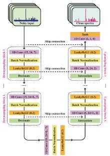
Fig 1
The architecture of LD-Net with L-layer downsampling and upsampling blocks for NMR spectroscopy denoising. 1D convolution (K, C, P) is used in which K, C, P denote the kernel size, channel number, the number of zero paddings, respectively. The original code scripts, trained models, as well as the example datasets would be freely available at https://github.com/HaolinZhan/LD-Net-for-NMR-spectroscopy-denoising."
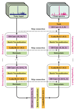
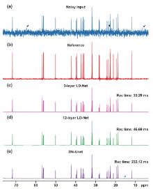
Fig 2
1D 13C NMR spectra of 0.1 mol∙L–1 cholesterol in CDCl3. (a) A noisy spectrum which is acquired with 4 scans. (b) The reference spectrum acquired with 512 scans. (c, d) The spectra using LD-Net denoising with the layers of 3 and 12, namely L = 3 (c) and L = 12 (d), respectively. (e) The spectra adopting DN-Unet denoising as a contrast. The spectral SNRs after denoising are enhanced by factors of 218 (c), 153 (d), and 277 (e) relative to the noisy input (a), respectively. All reconstruction times were counted on the identical computing hardware."
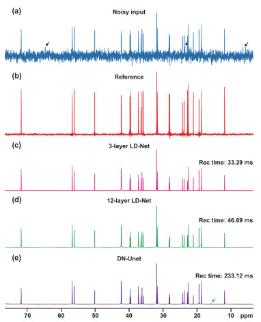
Table 1
Performance comparison between different methods for NMR spectroscopy denoising a."
| Methods | Training times (h) | Model sizes (MB) | Parameters (MB) | Reconstruction times (ms) | SNR enhancements | RMSDs (e-4) | R2 (%) | Error rates b |
| LD-Net (L = 3) | 5.98 | 0.82 | 0.20 | 32.3 ± 2.50 | 139.4 ± 46.47 | 9.2 ± 2.47 | 99.85 ± 0.13 | 0% |
| LD-Net (L = 12) | 4.32 | 38.76 | 10.13 | 46.1 ± 1.84 | 140.7 ± 16.05 | 8.4 ± 2.27 | 99.86 ± 0.13 | 0.5% |
| DN-Unet | 11.90 | 568.30 | 148.93 | 225.2 ± 12.11 | 238.1 ± 38.56 | 8.4 ± 2.28 | 99.85 ± 0.12 | 0.5% |
| Wavenet | 46.34 | 8.63 | 2.24 | 130.1 ± 5.36 | 97.1±40.87 | 9.3 ± 2.43 | 99.86 ± 0.12 | 0.5% |
| U-Net | 1.55 | 56.50 | 14.79 | 33.3 ± 1.01 | 135.1±47.28 | 11.2 ± 2.92 | 99.86 ± 0.11 | 14% |
| CoSeM | – | 3.3e5 ± 1.58e4 | 3.2 ± 0.70 c | 63.9 ± 0.042 | 99.51±0.27 | 0% | ||
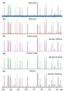
Fig 3
1D 13C NMR spectra of a mixture containing 0.5 mol∙L–1 quinine and 0.05 mol∙L–1 menthol in CDCl3. (a) A noisy spectrum acquired with 4 scans. (b) The reference spectrum acquired with 512 scans. (c, d) The spectra using LD-Net denoising with the layers of 3 and 12, namely L = 3 (c) and L = 12 (d), respectively. (e) The spectra using DN-Unet denoising as a contrast."
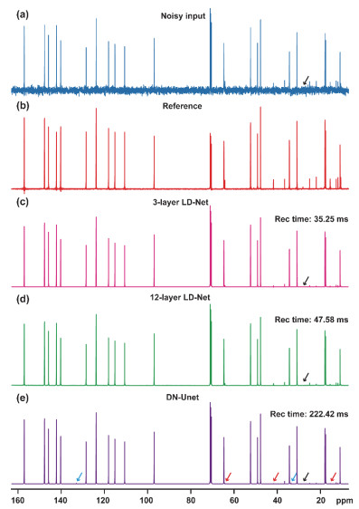
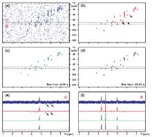
Fig 4
2D 1H-13C HSQC spectra of 0.2 mol∙L-1 azithromycin in CDCl3. (a) A noisy input acquired with 4 scans and additional Gaussian white noise with standard deviations of 0.02 is added. (b) 2D spectrum collected with 32 scans as a reference. (c, d) 2D spectra after 12-layer LD-Net denoising (c) and DN-Unet denoising (d). (e, f) 1D slices for each of 2D spectra from the indicated regions. The 2D contour levels for panels c and d remain identical for a fair comparison, and panels a and b are contoured with the smaller levels for the better visualization. The black arrows denote the peaks with negative phases in panels b and e."
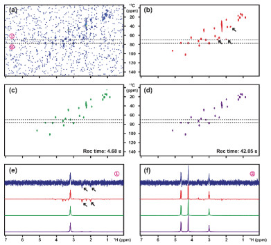
Fig 5
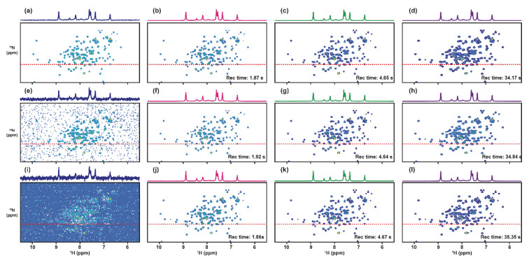
2D 1H-15N HSQC spectra of 0.25 mmol∙L–1 15N-labeled BRM bromodomain. (a, e, i) Noisy inputs acquired with 8 scans (a) and adding random Gaussian white noise with standard deviations of 0.02 (e) and 0.03 (f). (b, f, j) 2D spectra after 3-layer LD-Net denoising on inputs of panels a, e, i, respectively. (c, g, k) 2D spectra after 12-layer LD-Net denoising on inputs of panels a, e, i, respectively. (d, h, l) 2D spectra after DN-Unet denoising on inputs of panels a, e, i, respectively. The 2D contour levels for all panels apart from the panel l remain identical for a fair comparison, and the panel l is contoured with a smaller level for the better visualization."
| 1 |
doi: 10.1021/acs.chemrev.1c00937 |
| 2 |
doi: 10.1021/jacs.2c05332 |
| 3 |
doi: 10.1002/mrm.29562 |
| 4 |
doi: 10.1016/S1872-2067(23)64526-7 |
| 5 |
doi: 10.1016/j.aca.2023.341682 |
| 6 |
doi: 10.1021/acs.jpclett.0c03549 |
| 7 |
doi: 10.3866/PKU.WHXB201912074 |
|
徐君; 邓风. 物理化学学报, 2020, 36(4), 1912074.
doi: 10.3866/PKU.WHXB201912074 |
|
| 8 |
doi: 10.3866/PKU.WHXB202212025 |
|
胡荣; 韦丽云; 鲜靖林; 房光钰; 吴植傲; 樊淼; 郭家越; 李青翔; 刘凯思; 姜会钰; 等. 物理化学学报, 2023, 39(9), 2212025.
doi: 10.3866/PKU.WHXB202212025 |
|
| 9 |
doi: 10.3866/PKU.WHXB202212060 |
|
何展军; 黄敏; 林铁军; 钟良枢. 物理化学学报, 2023, 39(9), 2212060.
doi: 10.3866/PKU.WHXB202212060 |
|
| 10 |
doi: 10.1016/j.jmr.2017.08.007 |
| 11 |
doi: 10.1021/jacs.0c00590 |
| 12 |
doi: 10.1016/j.pnmrs.2005.03.001 |
| 13 |
doi: 10.1016/j.jmr.2016.03.006 |
| 14 |
doi: 10.1073/pnas.1909921117 |
| 15 |
doi: 10.1126/sciadv.ade2113 |
| 16 |
doi: 10.3866/PKU.WHXB201906018 |
|
庞振峰; 管晗曦; 高李娜; 曹伟成; 尹竟琳; 孔学谦. 物理化学学报, 2020, 36(4), 1906018.
doi: 10.3866/PKU.WHXB201906018 |
|
| 17 |
doi: 10.1016/j.pnmrs.2021.04.002 |
| 18 |
doi: 10.1002/anie.202203957 |
| 19 |
doi: 10.1038/s41467-022-28304-w |
| 20 |
doi: 10.1021/jacs.0c00807 |
| 21 |
doi: 10.1016/j.pnmrs.2020.11.002 |
| 22 |
doi: 10.1126/sciadv.abc8180 |
| 23 |
doi: 10.1016/j.pnmrs.2012.03.001 |
| 24 |
doi: 10.1021/jacs.9b10094 |
| 25 |
doi: 10.1016/j.pnmrs.2019.05.005 |
| 26 |
doi: 10.1016/j.jmr.2022.107187 |
| 27 |
doi: 10.1109/tim.2021.3109743 |
| 28 |
doi: 10.1021/ac303726p |
| 29 |
doi: 10.1021/acs.jpca.9b04437 |
| 30 |
doi: 10.1002/mrm.28654 |
| 31 |
doi: 10.1038/nature14539 |
| 32 |
doi: 10.1038/s41467-023-39581-4 |
| 33 |
Wang, W. L.; Ma, L. H.; Maletic-Savatic, M.; Liu, Z. D. NMRQNet: a deep learning approach for automatic identification and quantification of metabolites using Nuclear Magnetic Resonance (NMR) in human plasma samples. bioRxiv [Preprint], 2023. Available Online: https://www.ncbi.nlm.nih.gov/pubmed/36909516 (accessed on Mar 2, 2023).
|
| 34 |
doi: 10.1002/anie.201908162 |
| 35 |
doi: 10.1021/acs.jpclett.2c00100 |
| 36 |
doi: 10.1007/s10858-021-00366-w |
| 37 |
doi: 10.1021/jacs.1c04010 |
| 38 |
doi: 10.1021/acs.analchem.3c00537 |
| 39 |
doi: 10.1002/mrm.27727 |
| 40 |
doi: 10.1109/tci.2023.3267623 |
| 41 |
doi: 10.1021/acs.analchem.0c03087 |
| 42 |
Ronneberger, O.; Fischer, P.; Brox, T. U-Net: Convolutional networks for biomedical image segmentation. In Medical Image Computing and Computer-Assisted Intervention – MICCAI 2015, 18th International Conference, Munich, Germany, Oct. 5–9, 2015; Navab, N., Hornegger, J., Wells, W. M., Frangi, A. F., Eds.; Springer Nature: Berlin, Germany, 2015; pp. 234–241.
|
| 43 |
Stoller, D.; Ewert, S.; Dixon, S. A multi-scale neural network for end-to-end audio source separation. arxiv [Preprint], 2018. Available Online: https://arxiv.org/abs/1806.03185 (accessed on Jun 8, 2018).
|
| 44 |
Macartney, C.; Weyde, T. Improved speech enhancement with the Wave-U-Net. arXiv[Preprint], 2018. Available Online: https://arxiv.org/abs/1811.11307 (accessed on Nov 27, 2018).
|
| 45 |
doi: 10.1002/anie.201707114 |
| 46 |
Rethage, D.; Pons, J.; Serra, X. A Wavenet for speech denoising. In ICASSP 2018–2018 IEEE International Conference on Acoustics, Speech and Signal Processing (ICASSP), Calgary, AB, Canada, Apr. 15–20, 2018; IEEE: New York, US, 2018; pp. 5069–5073.
|
| 47 |
doi: 10.1016/j.pnmrs.2015.02.002 |
| 48 |
doi: 10.1021/acs.jpclett.9b03092 |
| 49 |
doi: 10.1021/acs.analchem.2c03678 |
| [1] | Hengrui Zhang, Xijun Xu, Xun-Lu Li, Xiangwen Gao. Applications of Generative Artificial Intelligence in Battery Research: Current Status and Prospects [J]. Acta Phys. -Chim. Sin., 2025, 41(10): 100115-. |
| [2] | QIU Xiao-Mei; LI Ling; WEI Xi-Lian; YIN Bao-Lin; SUN De-Zhi. A Study on Complexation between α-cyclodextrin and Bis-quaternary Ammonium Surfactants [J]. Acta Phys. -Chim. Sin., 2005, 21(12): 1415-1418. |
|
||