物理化学学报 >> 2025, Vol. 41 >> Issue (5): 100042.doi: 10.1016/j.actphy.2024.100042
高效靶向示踪钙钛矿纳米系统光电增效抗肿瘤
李坚1,2,3,†, 张毓2,†, 闫融融1,4,†, 孙开媛5, 刘晓晴1, 梁子尚1,4, 焦忆楠4, 卜晖2,*( ), 陈欣1,6,*(), 赵晋津1,*(), 施剑林7
- 1 河北师范大学化学与材料科学学院, 河北省能量转换材料与器件技术创新中心, 薄膜太阳能电池材料与器件工程研究中心, 河北省无机纳米材料重点实验室, 石家庄 050024
2 河北医科大学第二医院神经内科, 临床神经病学教育部重点实验室, 河北省神经病学重点实验室, 石家庄 050051
3 衡水市人民医院全科医学科, 河北衡水 053000
4 石家庄铁道大学材料科学与工程学院, 石家庄 050043
5 北京航空航天大学生物与医学工程学院, 北京 100191
6 邢台市人民医院神经内科, 邢台市神经病学重点实验室, 河北邢台 054001
7 中国科学院上海硅酸盐研究所, 上海 200050
-
收稿日期:2024-10-18修回日期:2024-11-29录用日期:2024-11-30发布日期:2025-04-18 -
通讯作者:Email: jinjinzhao2012@163.com (赵晋津) Email: jinjinzhao2012@163.com (Jinjin Zhao)26500825@hebmu.edu.cn (卜晖)xinc2019@126.com (陈欣) -
作者简介:†These authors contributed equally.
-
基金资助:国家自然科学基金(U2130128); 河北省自然科学基金-燕赵青年科学家专项(B2023205040); 河北省自然科学基金-京津冀基础研究合作专项(H2022205047); 河北省自然科学基金-京津冀基础研究合作专项(22JCZXJC00060); 河北省自然科学基金-京津冀基础研究合作专项(E3B33911DF); 中央引导地方科技发展资金项目(236Z7753G); 中央引导地方科技发展资金项目(246Z7755G); 河北省高等学校科学研究项目基础研究重点培育专项(JCZX2025007); 河北省科技厅引智与人才培养专项基金; 河北省创新能力提升计划(22567604H); 河北师范大学博士科研启动基金(L2023B18); 大学生创新创业训练计划项目(S202410094046)
Highly Efficient, Targeted, and Traceable Perovskite Nanocrystals for Photoelectrocatalytic Oncotherapy
Jian Li1,2,3, Yu Zhang2, Rongrong Yan1,4, Kaiyuan Sun5, Xiaoqing Liu1, Zishang Liang1,4, Yinan Jiao4, Hui Bu2,*(), Xin Chen1,6,*(), Jinjin Zhao1,*(), Jianlin Shi7
- 1 College of Chemistry and Materials Science, Hebei Technology Innovation Center for Energy Conversion Materials and Devices, Hebei Key Laboratory of Inorganic Nanomaterials, Engineering Research Center of Thin Film Solar Cell Materials and Devices, Hebei Province, Hebei Normal University, Shijiazhuang 050024, China
2 Department of Neurology, The Second Hospital of Hebei Medical University, Key Laboratory of Clinical Neurology (Hebei Medical University), Ministry of Education, Neurological Laboratory of Hebei Province, Shijiazhuang 050051, China
3 General Practice Department, Hengshui People's Hospital, Hengshui 053000, Hebei Province, China
4 School of Materials Science and Engineering, Shijiazhuang Tiedao University, Shijiazhuang 050043, China
5 School of Biological Science and Medical Engineering, Beihang University, Beijing 100191, China
6 Department of Neurology, Xingtai People's Hospital, Xingtai Key Laboratory of Neurology, Xingtai 054001, Hebei Province, China
7 Shanghai Institute of Ceramics, Chinese Academy of Sciences, Shanghai 200050, China
-
Received:2024-10-18Revised:2024-11-29Accepted:2024-11-30Published:2025-04-18 -
Contact:26500825@hebmu.edu.cn (Hui Bu)xinc2019@126.com (Xin Chen) -
Supported by:the National Natural Science Foundation of China(U2130128); the Yanzhao Young Scientist Project from Hebei Natural Science Foundation(B2023205040); the Basic Research Cooperation Special Foundation of Beijing-Tianjin-Hebei Region from Hebei Natural Science Foundation(H2022205047); the Basic Research Cooperation Special Foundation of Beijing-Tianjin-Hebei Region from Hebei Natural Science Foundation(22JCZXJC00060); the Basic Research Cooperation Special Foundation of Beijing-Tianjin-Hebei Region from Hebei Natural Science Foundation(E3B33911DF); the Central Government Guiding Local Science and Technology Development Project(236Z7753G); the Central Government Guiding Local Science and Technology Development Project(246Z7755G); Key Cultivation Special Project for Basic Research from Hebei Education Department(JCZX2025007); Introducing Intelligence and Talent Cultivation Special Fundation from Hebei Provincial Department of Science and Technology; the Innovation Capability Improvement Plan Project of Hebei Province(22567604H); the Ph.D Scientific Research Start-up Fund of Hebei Normal University(L2023B18); the College student's innovation and entrepreneurship training plan program(S202410094046)

摘要:
金属卤化物钙钛矿由于其多维度的晶体结构和优良的荧光成像/示踪及光电转换性质,使其成为一种非常具有前瞻性的光电增效治疗肿瘤材料。然而,传统卤化物钙钛矿纳米晶的水稳定性问题,限制了其应用于生物成像和光电增效肿瘤治疗的药物递送纳米系统研究。本文将甲氨蝶呤-壳聚糖-叶酸(MTX-CS-FA)成功与钙钛矿纳米晶体CsSn0.5Pb0.5Br3 (PeNCs)铆钉连接,制备出了可以在水中稳定228 d且发绿光的PeNCs@MTX-CS-FA纳米载药体系。在可见光照射下,新型PeNCs@MTX-CS-FA纳米载药体系增效抗肿瘤治疗原理:钙钛矿纳米晶体产生电子和活性氧(ROS);钙钛矿光生空穴耗竭过表达的谷胱甘肽(GSH);甲氨蝶呤(MTX)抑制二氢叶酸还原酶(DHFR)活性,导致肿瘤细胞的脂质过氧化,上述三点共同作用抑制肿瘤细胞的增殖、促进肿瘤细胞凋亡。在动物体内实验中,采用小鼠移植肿瘤模型,累积用药量达2.4 mg PeNCs@MTX-CS-FA纳米载药系统时,肿瘤体积减少了约63.68%和肿瘤重量下降了约63.26%。通过生物安全性评估实验证实,在治疗剂量下,小鼠肝、肾等器官功能正常,说明纳米体系具有良好的生物安全性,并且研究发现钙钛矿纳米颗粒经小鼠肠道排出,小鼠粪便呈现出与原始钙钛矿晶体相同的绿色荧光,金属卤化物钙钛矿纳米载药体系在生物成像和光电催化化疗方面呈现优异的增效抗肿瘤治疗效果。
引用本文
李坚, 张毓, 闫融融, 孙开媛, 刘晓晴, 梁子尚, 焦忆楠, 卜晖, 陈欣, 赵晋津, 施剑林. 高效靶向示踪钙钛矿纳米系统光电增效抗肿瘤[J]. 物理化学学报, 2025, 41(5), 100042. doi: 10.1016/j.actphy.2024.100042
Jian Li, Yu Zhang, Rongrong Yan, Kaiyuan Sun, Xiaoqing Liu, Zishang Liang, Yinan Jiao, Hui Bu, Xin Chen, Jinjin Zhao, Jianlin Shi. Highly Efficient, Targeted, and Traceable Perovskite Nanocrystals for Photoelectrocatalytic Oncotherapy[J]. Acta Phys. -Chim. Sin. 2025, 41(5), 100042. doi: 10.1016/j.actphy.2024.100042
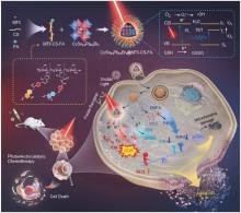
图1
"
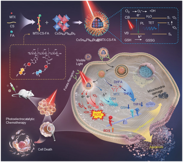
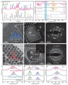
图2
"
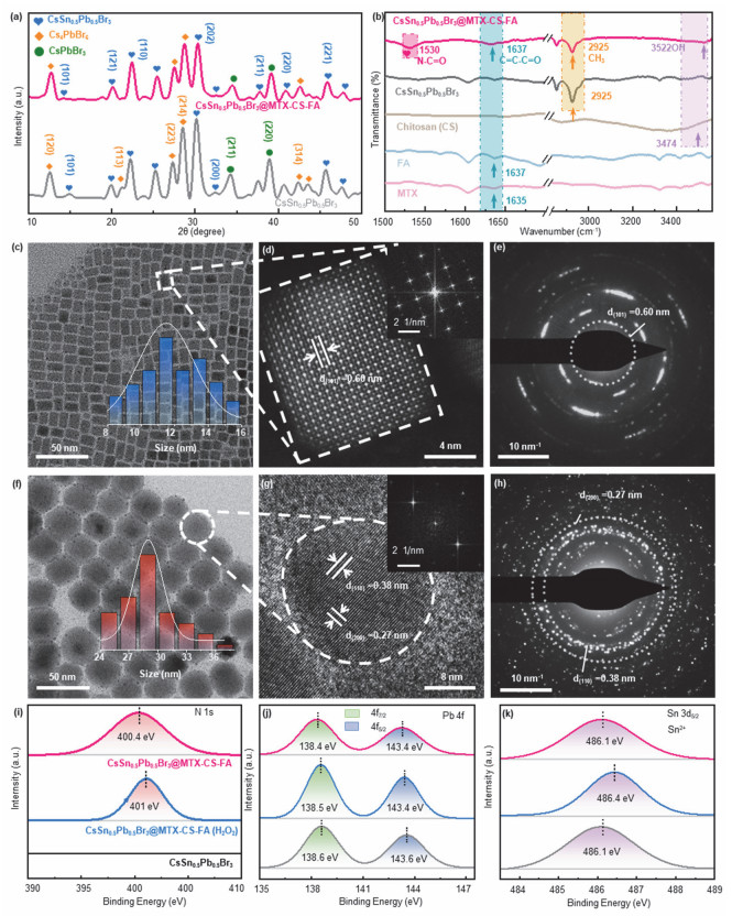
图3
"
图4
"
图5
"
图6
"
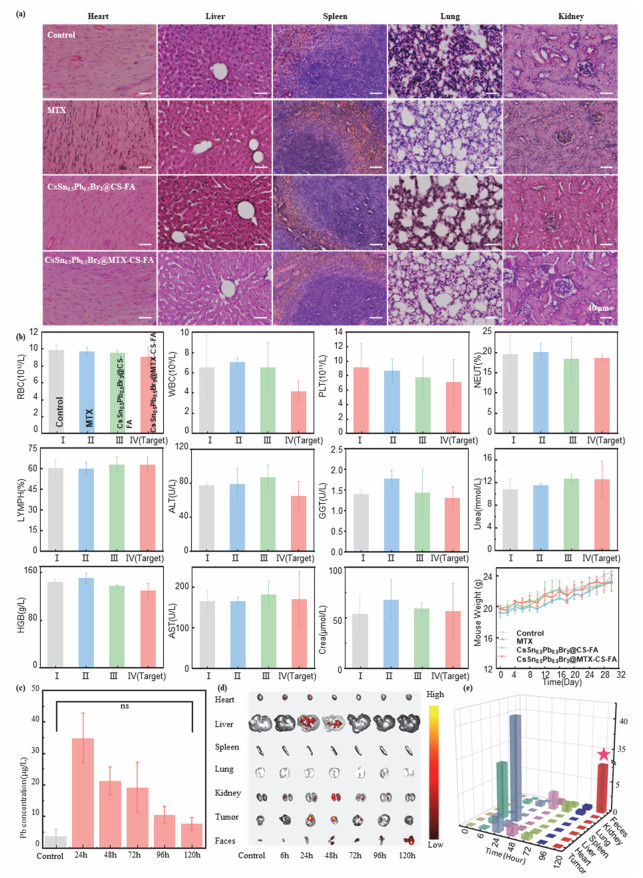
图7
"
| 1 |
doi: 10.1038/35104607 |
| 2 |
doi: 10.1038/nature12340 |
| 3 |
doi: 10.1126/sciadv.ade9044 |
| 4 |
doi: 10.1038/s41586-023-06514-6 |
| 5 |
doi: 10.1038/s41467-021-27640-7 |
| 6 |
doi: 10.1021/jacs.2c12942 |
| 7 |
doi: 10.1002/adma.202208817 |
| 8 |
doi: 10.1038/s41566-021-00931-7 |
| 9 |
doi: 10.1038/s41467-023-35837-1 |
| 10 |
doi: 10.1016/j.nanoen.2022.108074 |
| 11 |
doi: 10.1016/j.scib.2017.08.022 |
| 12 |
doi: 10.1039/c8ta05282d |
| 13 |
doi: 10.1002/adma.202206684 |
| 14 |
doi: 10.1021/ic401215x |
| 15 |
doi: 10.1126/science.aaa5760 |
| 16 |
doi: 10.1002/adma.201806671 |
| 17 |
doi: 10.1126/sciadv.abg8458 |
| 18 |
doi: 10.1038/s41586-022-05486-3 |
| 19 |
doi: 10.1002/smll.202204763 |
| 20 |
doi: 10.1002/smll.202310238 |
| 21 |
doi: 10.1007/s12598-022-02222-8 |
| 22 |
doi: 10.1126/science.adf3349 |
| 23 |
doi: 10.1126/science.adi4107 |
| 24 |
doi: 10.1038/s41586-023-06006-7 |
| 25 |
doi: 10.1038/s41586-023-06396-8 |
| 26 |
doi: 10.1021/acsphotonics.3c00042 |
| 27 |
doi: 10.1002/adfm.202102334 |
| 28 |
doi: 10.1016/j.ccr.2021.214313 |
| 29 |
doi: 10.1186/s40580-023-00366-6 |
| 30 |
doi: 10.1038/s41467-019-13951-3 |
| 31 |
doi: 10.1021/jacs.0c04583 |
| 32 |
doi: 10.1002/anie.202208241 |
| 33 |
doi: 10.1016/j.chempr.2022.03.003 |
| 34 |
doi: 10.1038/ncomms12555 |
| 35 |
doi: 10.1038/s41467-020-15016-2 |
| 36 |
doi: 10.1007/s42765-022-00207-x |
| 37 |
doi: 10.1021/acsanm.3c05605 |
| 38 |
doi: 10.1016/j.nanoen.2019.03.053 |
| 39 |
doi: 10.1002/adfm.202202951 |
| 40 |
doi: 10.1016/j.jpowsour.2016.11.046 |
| 41 |
doi: 10.1002/adma.202203430 |
| 42 |
doi: 10.1002/lpor.202400367 |
| 43 |
doi: 10.1016/j.msec.2020.110807 |
| 44 |
doi: 10.1021/acsami.8b00947 |
| 45 |
doi: 10.1021/acsanm.2c05482 |
| 46 |
doi: 10.1016/j.jcis.2018.06.024 |
| 47 |
doi: 10.1038/s41565-022-01163-8 |
| 48 |
doi: 10.1016/j.cej.2023.143457 |
| 49 |
doi: 10.1002/smll.202300101 |
| 50 |
doi: 10.1002/ange.202006169 |
| 51 |
doi: 10.1002/smll.202307972 |
| 52 |
doi: 10.1002/adma.202005888 |
| 53 |
doi: 10.1038/s41467-022-32050-4 |
| 54 |
doi: 10.1038/s41467-022-33474-8 |
| 55 |
doi: 10.1002/adfm.202213364 |
| 56 |
doi: 10.1002/adfm.202215244 |
| 57 |
doi: 10.1002/anie.202103721 |
| 58 |
doi: 10.1021/jacs.8b04400 |
| 59 |
doi: 10.1002/anie.202007786 |
| 60 |
doi: 10.1016/j.carres.2021.108275 |
| 61 |
doi: 10.1021/mp400269z |
| 62 |
doi: 10.1021/acsnano.2c03752 |
| 63 |
doi: 10.1016/j.jconrel.2023.05.040 |
| 64 |
doi: 10.1016/j.cmet.2020.10.011 |
| 65 |
doi: 10.1021/jacsau.0c00038 |
| 66 |
doi: 10.1038/s41586-022-04961-1 |
| 67 |
doi: 10.1038/s41586-023-05938-4 |
| 68 |
doi: 10.1002/advs.202203749 |
| 69 |
doi: 10.1021/acsenergylett.2c02262 |
| 70 |
doi: 10.1021/jacs.7b08628 |
| 71 |
doi: 10.1021/acsami.0c20285 |
| 72 |
doi: 10.1021/acsmaterialslett.0c00596 |
| 73 |
doi: 10.1021/jacs.5b13470 |
| 74 |
doi: 10.1039/D3TA02909C |
| 75 |
http://www.science-and-fun.de/tools/ (accessed on Oct 3, 2024).
|
| 76 |
doi: 10.3390/ph15020149 |
| 77 |
doi: 10.1016/j.xphs.2021.10.034 |
| 78 |
doi: 10.1039/d0nr08325a |
| 79 |
doi: 10.1016/j.cej.2021.131950 |
| 80 |
doi: 10.1002/adom.202202673 |
| 81 |
doi: 10.1016/j.apcatb.2016.04.016 |
| 82 |
doi: 10.1088/1757-899x/490/2/022067 |
| 83 |
doi: 10.1002/solr.201900457 |
| 84 |
doi: 10.1016/j.surfrep.2010.10.001 |
| 85 |
Chen, X.; Lai, J.; Shen, Y.; Chen, Q.; Chen, L. 2018, 30 (48), 1802490. doi: 10.1002/adma.201802490
|
| 86 |
doi: 10.1039/D2TA08349C |
| 87 |
Yuan, Y.; Li, T.; Wang, Q.; Xing, J.; Gruverman, A.; Huang, J. 2017, 3 (3), e1602164. doi: 10.1126/sciadv.1602164
|
| 88 |
doi: 10.1002/anie.202203088 |
| 89 |
doi: 10.1038/s41467-022-34036-8 |
| 90 |
doi: 10.1038/nm.4232 |
| 91 |
doi: 10.1002/adma.202209589 |
| 92 |
doi: 10.1016/j.biomaterials.2021.121110 |
| 93 |
doi: 10.1002/smll.202303365 |
| 94 |
doi: 10.1038/s41467-022-31799-y |
| 95 |
doi: 10.1016/j.ccell.2018.12.009 |
| 96 |
doi: 10.1039/d0cs00718h |
| 97 |
doi: 10.1093/nsr/nwab139 |
| 98 |
doi: 10.1038/s41568-021-00435-0 |
| 99 |
doi: 10.1021/nn5061404 |
| 100 |
doi: 10.1016/j.biomaterials.2022.121724 |
| 101 |
doi: 10.1021/acsabm.0c00171 |
| 102 |
doi: 10.1002/adma.202200871 |
| 103 |
doi: 10.1016/j.colsurfb.2023.113308 |
| 104 |
doi: 10.1016/j.ijbiomac.2022.07.185 |
| 105 |
doi: 10.1186/s12645-023-00162-1 |
| 106 |
doi: 10.1021/acs.molpharmaceut.2c01035 |
| 107 |
doi: 10.1016/j.apsb.2021.09.024 |
| 108 |
doi: 10.1039/D2BM00883A |
| 109 |
doi: 10.1038/s41467-022-32349-2 |
| 110 |
doi: 10.1016/j.chempr.2020.06.024 |
| 111 |
doi: 10.1002/adma.202008065 |
| 112 |
doi: 10.1016/j.cej.2021.129178 |
| 113 |
doi: 10.1016/j.cej.2022.135373 |
| 114 |
https://stacks.cdc.gov/view/cdc/37586 (accessed on Oct 3, 2024).
|
| 115 |
doi: 10.1016/j.phrs.2022.106632 |
| [1] | 邵仁义, 库拉姆·阿巴斯, 弗拉基米尔·尤里耶维奇·奥西波夫, 朱海梅, 李远, 乌萨马, 毕红. 源自绿萝叶提取物的红光碳点制备和生物成像应用[J]. 物理化学学报, 2026, 42(2): 100134 - . |
| [2] | 胡超,穆野,李明宇,邱介山. 纳米碳点的制备与应用研究进展[J]. 物理化学学报, 2019, 35(6): 572 -590 . |
| [3] | 宋雪娇,刘庄. 纳米技术在物理刺激诱导肿瘤治疗中的应用探索[J]. 物理化学学报, 2018, 34(2): 123 -139 . |
| [4] | 陈凡,王中跃,张艳艳,余柯涵,翁丽星,韦玮. 聚丙烯酸功能化高效发光的La1-xEuxF3纳米晶合成及细胞成像[J]. 物理化学学报, 2017, 33(7): 1446 -1452 . |
|
||