Acta Phys. -Chim. Sin. ›› 2020, Vol. 36 ›› Issue (12): 2007066.doi: 10.3866/PKU.WHXB202007066
Special Issue: Neural Interfaces
• REVIEW • Previous Articles Next Articles
Carbon-based Nanomaterials for Neural Electrode Technology
Yang Liu1, Xiaojie Duan1,2,*( )
)
- 1 Department of Biomedical Engineering, College of Engineering, Peking University, Beijing 100871, China
2 Academy for Advanced Interdisciplinary Studies, Peking University, Beijing 100871, China
-
Received:2020-07-25Accepted:2020-08-23Published:2020-08-27 -
Contact:Xiaojie Duan E-mail:xjduan@pku.edu.cn -
Supported by:the National Natural Science Foundation of China(21972005);the National Natural Science Foundation of China(91648207);the National Natural Science Foundation of China(81771821);the National Key Basic Research Program of China(2016YFA0200103);the Beijing Graphene Innovation Program(Z191100000819001)

Cite this article
Yang Liu, Xiaojie Duan. Carbon-based Nanomaterials for Neural Electrode Technology[J]. Acta Phys. -Chim. Sin. 2020, 36(12), 2007066. doi: 10.3866/PKU.WHXB202007066
share this article
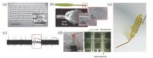
Fig 1
Soft graphene electrodes. (a) Tilt SEM image of a 64-spot porous graphene array. Scale bar, 1 mm. The inset is the SEM image of an individual electrode active site. Scale bar: 100 μm9. Adapted from Ref.9. (b and c) Laser ablation of liquid crystal graphene oxide (LCGO) electrodes and neural activity of feline visual cortex recorded within 20 s of LCGO electrode implantation 12. Adapted from Ref.12. (d) (left) Photograph and (right) optical microscope images of the electrode array consisting of electrode sites based on Au film, ZnO nanowires and poly(3, 4-ethylenedioxythiophene (PEDOT), and interconnection line based on Au/graphene 38. Scale bars, (left) 10 mm and (right) 450 μm. Adapted from Ref.38. (e) Image of 4 × 4 graphene transistor array 42. Adapted from Ref.42. "
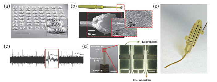
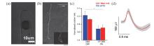
Fig 2
Soft carbon nanotubes (CNTs) electrodes. (a) SEM image of a CNT:poly(sodium 4-styrenesulfonate) (PSS)/poly(vinyl alcohol) (PVA) composite electrode with a width of 10 μm 11. Adapted from Ref.11. (b) Two-channel CNT fiber microelectrodes; inset shows a close view of the active site 49. Scale bar, 200 μm. (c) Results of the methamphetamine rotation test 49. (d) Mean waveforms of single units averaged over multiple 40 sample traces detected by CNT fiber electrodes (the shaded area depicts the mean ± SD) 49. Adapted from Ref.49. "
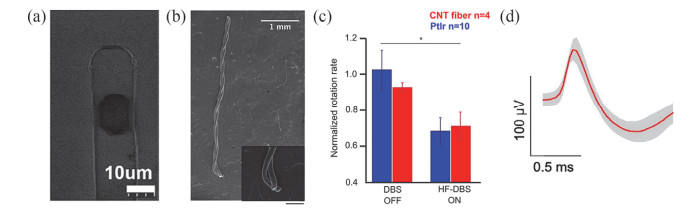
Fig 3
Chronic neural recording with soft CNT fiber electrodes 33. (a) Four-channel CNT fiber microelectrode array. Scale bar, 5 mm. Inset, SEM image of a CNT fiber electrode tip. Scale bar, 10 μm. (b) Mean waveforms of units detected and isolated from day 1 to day 117 postimplantation with an ~15 μm diameter CNT fiber electrodes. Adapted from Ref.33."
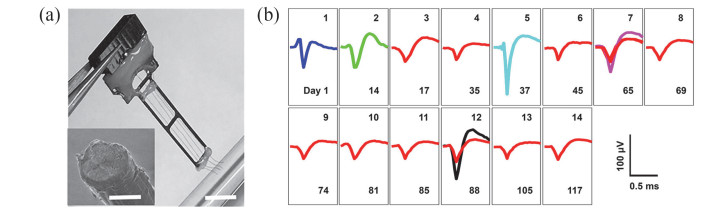
Fig 4
Schematics of the two-layer PDMS microfluidic device assisting insertion of flexible CNTf microelectrodes 56. Adapted from Ref.56. "
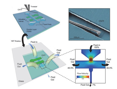
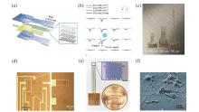
Fig 5
Transparent graphene electrodes. (a) Diagram of CLEAR device construction 23. Adapted from Ref.23. (b) Optical evoked potentials recorded by CLEAR device 23. x-scale bars represent 50 ms, y-scale bars represent 100 μV. Adapted from Ref.23. (c) Three different types of transparent graphene multielectrode arrays with different electrode site diameters (100, 150, 200 μm) 58. Adapted from Ref.58. (d) Microscope image of transparent graphene 8-electrode array 14. Adapted from Ref.14. (e) Transparent graphene microelectrode arrays with 16 electrode sites. Each electrode is a 100 μm×100 μm square 26. Scale bar, 500 μm. Adapted from Ref.26. (f) FE-SEM images of the fixed rat cortical neuron cultured on transparent graphene-vertically aligned carbon nanotube hybrid electrodes (TGVH) at 7 days in vitro (DIV) 59. Adapted from Ref.59."
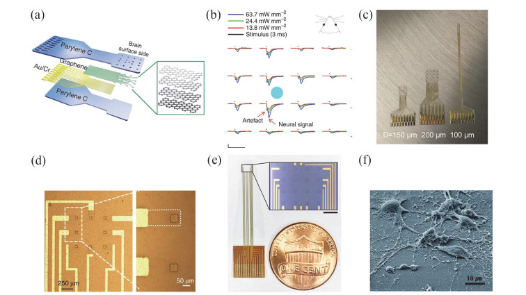
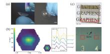
Fig 6
Soft transparent graphene contact lens electrodes (GRACE) 61. (a) Photographs of a GRACE device. Scale bar, 3 mm. Image in the inset demonstrates the high softness of the GRACE. (b) The mfERG responses recorded with a GRACE device. (c) Top, soft and transparent graphene electrode array. Scale bar, 5 mm. Bottom, optical microscopy image showing some of the graphene electrode sites and traces. Scale bar, 150 μm. Adapted from Ref.61. "

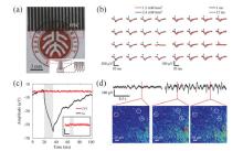
Fig 7
Transparent carbon nanotubes electrodes 63. (a) Stretchable transparent CNT electrode array, (b) Light-evoked potentials recorded by the stretchable transparent CNT electrode array under different stimulus intensity and duration. (c) The light-induced artifacts in CNT and Au electrode recording with a recording-site size of 100 μm × 100 μm tested in saline. (d) ECoG recording from a stretchable transparent CNT electrode and two-photon calcium images of the brain region underneath the recording site of the electrode used to record the top traces. Adapted from Ref.63. "
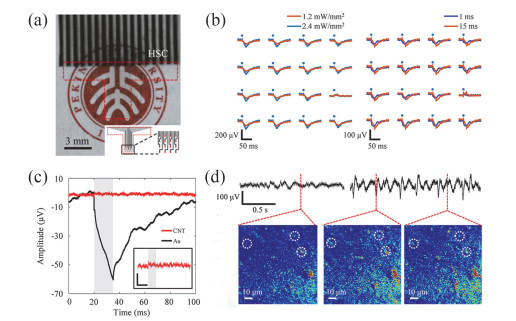
Fig 8
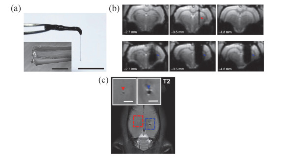
MRI compatible neural electrodes made from carbon nanomaterials. (a) GF bipolar microelectrode assembly 32. Scale bar, 1 cm; inset, 100 μm. Adapted from Ref.32. (b) Representative three serial coronal scans from rostral (left) to caudal (right) of EPI images from rat brains implanted with a GF (top) and PtIr (bottom) bipolar microelectrode, with the middle images depicting the electrode implant sites 32. Adapted from Ref.32. (c) Horizontal sections of the T2-weighted images of a rat implanted contralaterally with a Parylene-C-insulated CNT fiber (red) and PtIr microwire (blue) 33. Scale bar, 1.5 mm (inset). Adapted from Ref.33. "
| 1 |
Quiroga R. Q. ; Reddy L. ; Kreiman G. ; Koch C. ; Fried I. Nature 2005, 435 (7045), 1102.
doi: 10.1038/nature03687 |
| 2 |
Jang A. I. ; Wittig J. H. ; Jr. ; Inati S. K. ; Zaghloul K. A. Curr. Biol. 2017, 27 (11), 1700.
doi: 10.1016/j.cub.2017.05.014 |
| 3 |
Lee K. Y. ; Ratte S. ; Prescott S. A. Elife 2019, 8, e49753.
doi: 10.7554/eLife.49753 |
| 4 |
Truccolo W. ; Donoghue J. A. ; Hochberg L. R. ; Eskandar E. N. ; Madsen J. R. ; Anderson W. S. ; Brown E. N. ; Halgren E. ; Cash S. S. Nat. Neurosci. 2011, 14 (5), 635.
doi: 10.1038/nn.2782 |
| 5 |
Fried I. ; Macdonald K. A. ; Wilson C. L. Neuron 1997, 18 (5), 753.
doi: 10.1016/S0896-6273(00)80315-3 |
| 6 |
Middlebrooks J. C. ; Bierer J. A. ; Snyder R. L. Curr. Opin. Neurobiol. 2005, 15 (4), 488.
doi: 10.1016/j.conb.2005.06.004 |
| 7 |
Benabid A. L. ; Chabardes S. ; Mitrofanis J. ; Pollak P. Lancet Neurol. 2009, 8 (1), 67.
doi: 10.1016/S1474-4422(08)70291-6 |
| 8 |
Wolter T. J. Pain Res. 2014, 7, 651.
doi: 10.2147/JPR.S37589 |
| 9 |
Lu Y. ; Lyu H. ; Richardson A. G. ; Lucas T. H. ; Kuzum D. Sci. Rep. 2016, 6, 33526.
doi: 10.1038/srep33526 |
| 10 |
Lacour S. P. ; Courtine G. ; Guck J. Nat. Rev. Mater. 2016, 1, 16063.
doi: 10.1038/natrevmats.2016.63 |
| 11 |
Zhang H. ; Patel P. R. ; Xie Z. ; Swanson S. D. ; Wang X. ; Kotov N. A. ACS Nano 2013, 7 (9), 7619.
doi: 10.1021/nn402074y |
| 12 |
Apollo N. V. ; Maturana M. I. ; Tong W. ; Nayagam D. A. X. ; Shivdasani M. N. ; Foroughi J. ; Wallace G. G. ; Prawer S. ; Ibbotson M. R. ; Garrett D. J. Adv. Funct. Mater. 2015, 25 (23), 3551.
doi: 10.1002/adfm.201500110 |
| 13 |
Wang M. ; Mi G. ; Shi D. ; Bassous N. ; Hickey D. ; Webster T. J. Adv. Funct. Mater. 2018, 28 (12), 1700905.
doi: 10.1002/adfm.201700905 |
| 14 |
Kuzum D. ; Takano H. ; Shim E. ; Reed J. C. ; Juul H. ; Richardson A. G. ; de Vries J. ; Bink H. ; Dichter M. A. ; Lucas T. H. ; et al Nat. Commun. 2014, 5, 5259.
doi: 10.1038/ncomms6259 |
| 15 |
Baranauskas G. ; Maggiolini E. ; Castagnola E. ; Ansaldo A. ; Mazzoni A. ; Angotzi G. N. ; Vato A. ; Ricci D. ; Panzeri S. ; Fadiga L. J. Neural Eng. 2011, 8 (6), 066013.
doi: 10.1088/1741-2560/8/6/066013 |
| 16 |
Cogan S. F. Annu. Rev. Biomed. Eng. 2008, 10, 275.
doi: 10.1146/annurev.bioeng.10.061807.160518 |
| 17 | Xie F. ; Xi Y. ; Xu Q. D. ; Liu J. Q. Acta Phys. -Chim. Sin. 2020, 36, 2003014. |
|
谢凡; 奚野; 徐庆达; 刘景全. 物理化学学报, 2020, 36, 2003014.
doi: 10.3866/PKU.WHXB202003014 |
|
| 18 |
Moffitt M. A. ; McIntyre C. C. Clin. Neurophysiol. 2005, 116 (9), 2240.
doi: 10.1016/j.clinph.2005.05.018 |
| 19 |
Frank J. A. ; Antonini M. J. ; Anikeeva P. Nat. Biotechnol. 2019, 37 (9), 1013.
doi: 10.1038/s41587-019-0198-8 |
| 20 |
Song E. ; Li J. ; Won S. M. ; Bai W. ; Rogers J. A. Nat. Mater. 2020, 19 (6), 590.
doi: 10.1038/s41563-020-0679-7 |
| 21 |
Yang X. ; Zhou T. ; Zwang T. J. ; Hong G. ; Zhao Y. ; Viveros R. D. ; Fu T. M. ; Gao T. ; Lieber C. M. Nat. Mater. 2019, 18 (5), 510.
doi: 10.1038/s41563-019-0292-9 |
| 22 |
Minev I. R. ; Musienko P. ; Hirsch A. ; Barraud Q. ; Wenger N. ; Moraud E. M. ; Gandar J. ; Capogrosso M. ; Milekovic T. ; Asboth L. ; et al Science 2015, 347 (6218), 159.
doi: 10.1126/science.1260318 |
| 23 |
Park D. W. ; Schendel A. A. ; Mikael S. ; Brodnick S. K. ; Richner T. J. ; Ness J. P. ; Hayat M. R. ; Atry F. ; Frye S. T. ; Pashaie R. ; et al Nat. Commun. 2014, 5, 5258.
doi: 10.1038/ncomms6258 |
| 24 |
Kim T. ; Cho M. ; Yu K. J. Materials 2018, 11 (7), 1163.
doi: 10.3390/ma11071163 |
| 25 |
Han X. ; Qian X. ; Bernstein J. G. ; Zhou H. H. ; Franzesi G. T. ; Stern P. ; Bronson R. T. ; Graybiel A. M. ; Desimone R. ; Boyden E. S. Neuron 2009, 62 (2), 191- 8.
doi: 10.1016/j.neuron.2009.03.011 |
| 26 |
Thunemann M. ; Lu Y. ; Liu X. ; Kilic K. ; Desjardins M. ; Vandenberghe M. ; Sadegh S. ; Saisan P. A. ; Cheng Q. ; Weldy K. L. ; et al Nat. Commun. 2018, 9, 2035.
doi: 10.1038/s41467-018-04457-5 |
| 27 |
Zhao S. ; Liu X. ; Xu Z. ; Ren H. ; Deng B. ; Tang M. ; Lu L. ; Fu X. ; Peng H. ; Liu Z. ; Duan X. Nano Lett. 2016, 16 (12), 7731.
doi: 10.1021/acs.nanolett.6b03829 |
| 28 |
Wang X. Y. ; Narita A. ; Müllen K. Nat. Rev. Chem. 2018, 2, 0100.
doi: 10.1038/s41570-017-0100 |
| 29 |
Zhang M. ; Atkinson K. R. ; Baughman R. H. Science 2004, 306 (5700), 1358.
doi: 10.1126/science.1104276 |
| 30 |
McCallum G. A. ; Sui X. ; Qiu C. ; Marmerstein J. ; Zheng Y. ; Eggers T. E. ; Hu C. ; Dai L. ; Durand D. M. Sci. Rep. 2017, 7 (1), 11723.
doi: 10.1038/s41598-017-10639-w |
| 31 |
Guo Y. ; Duan W. ; Ma C. ; Jiang C. ; Xie Y. ; Hao H. ; Wang R. ; Li L. Biomed. Eng. Online 2015, 14, 118.
doi: 10.1186/s12938-015-0113-6 |
| 32 |
Zhao S. ; Li G. ; Tong C. ; Chen W. ; Wang P. ; Dai J. ; Fu X. ; Xu Z. ; Liu X. ; Lu L. ; et al Nat. Commun. 2020, 11, 1788.
doi: 10.1038/s41467-020-15570-9 |
| 33 |
Lu L. ; Fu X. ; Liew Y. ; Zhang Y. ; Zhao S. ; Xu Z. ; Zhao J. ; Li D. ; Li Q. ; Stanley G. B. ; Duan X. Nano Lett. 2019, 19 (3), 1577.
doi: 10.1021/acs.nanolett.8b04456 |
| 34 |
Xiao T. ; Wang Y. ; Wei H. ; Yu P. ; Jiang Y. ; Mao L. Angew. Chem., Int. Ed. 2019, 58 (20), 6616.
doi: 10.1002/anie.201901035 |
| 35 |
Geim A. K. ; Novoselov K. S. Nat. Mater. 2007, 6, 183.
doi: 10.1038/nmat1849 |
| 36 |
Lee C. ; Wei X. ; Kysar J. W. ; Hone J. S. Science 2008, 321 (5887), 385.
doi: 10.1126/science.1157996 |
| 37 |
Bae S. ; Kim H. ; Lee Y. ; Xu X. ; Park J. S. ; Zheng Y. ; Balakrishnan J. ; Lei T. ; Kim H. R. ; Song Y. I. ; et al Nat. Nanotechnol. 2010, 5, 574.
doi: 10.1038/nnano.2010.132 |
| 38 |
Ryu M. ; Yang J. H. ; Ahn Y. ; Sim M. ; Lee K. H. ; Kim K. ; Lee T. ; Yoo S. J. ; Kim S. Y. ; Moon C. ; et al ACS Appl. Mater. Interfaces 2017, 9 (12), 10577.
doi: 10.1021/acsami.7b02975 |
| 39 |
Blaschke B. M. ; Lottner M. ; Drieschner S. ; Calia A. B. ; Stoiber K. ; Rousseau L. ; Lissourges G. ; Garrido J. A. 2D Mater. 2016, 3 (2), 25007.
doi: 10.1088/2053-1583/3/2/025007 |
| 40 |
Hess L. H. ; Jansen M. ; Maybeck V. ; Hauf M. V. ; Seifert M. ; Stutzmann M. ; Sharp I. D. ; Offenhausser A. ; Garrido J. A. Adv. Mater. 2011, 23 (43), 5045.
doi: 10.1016/j.carbon.2018.11.026 |
| 41 |
Rauti R. ; Musto M. ; Bosi S. ; Prato M. ; Ballerini L. Carbon 2019, 143, 430.
doi: 10.1016/j.carbon.2018.11.026 |
| 42 |
Blaschke B. M. ; Tort-Colet N. ; Guimerà-Brunet A. ; Weinert J. ; Rousseau L. ; Heimann A. ; Drieschner S. ; Kempski O. ; Villa R. ; Sanchez-Vives M. V. ; Garrido J. A. 2D Mater. 2017, 4, 025040.
doi: 10.1088/2053-1583/aa5eff |
| 43 |
Peigney A. ; Laurent C. ; Flahaut E. ; Bacsa R. R. ; Rousset A. Carbon 2001, 39 (4), 507.
doi: 10.1016/S0008-6223(00)00155-X |
| 44 |
Shoval A. ; Adams C. ; David-Pur M. ; Shein M. ; Hanein Y. ; Sernagor E. Front. Neuroeng. 2009, 2, 4.
doi: 10.3389/neuro.16.004.2009 |
| 45 |
Kim H. i. ; Wang M. ; Lee S. K. ; Kang J. ; Nam J. D. ; Ci L. ; Suhr J. Sci. Rep. 2017, 7, 9512.
doi: 10.1038/s41598-017-10279-0 |
| 46 |
Wang L. ; Xie S. ; Wang Z. ; Liu F. ; Yang Y. ; Tang C. ; Wu X. ; Liu P. ; Li Y. ; Saiyin H. ; et al Nat. Biomed. Eng. 2020, 4, 159.
doi: 10.1038/s41551-019-0462-8 |
| 47 |
Yu X. ; Su J. Y. ; Guo J. Y. ; Zhang X. H. ; Li R. H. ; Chai X. Y. ; Chen Y. ; Zhang D. G. ; Wang J. G. ; Sui X. H. ; Durand D. M. J. Neurosci. Methods 2019, 328, 108450.
doi: 10.1016/j.jneumeth.2019.108450 |
| 48 |
Sanders J. E. ; Stiles C. E. ; Hayes C. L. J. Biomed. Mater. Res. 2000, 52 (1), 231.
doi: 10.1002/1097-4636(200010)52:1<231::AID-JBM29>3.0.CO;2-E |
| 49 |
Vitale F. ; Summerson S. R. ; Aazhang B. ; Kemere C. ; Pasquali M. ACS Nano 2015, 9 (4), 4465.
doi: 10.1021/acsnano.5b01060 |
| 50 |
Seo K. J. ; Artoni P. ; Qiang Y. ; Zhong Y. ; Han X. ; Shi Z. ; Yao W. ; Fagiolini M. ; Fang H. Adv. Biosyst. 2019, 3 (3), 1800276.
doi: 10.1002/adbi.201800276 |
| 51 |
Lind G. ; Linsmeier C. E. ; Thelin J. ; Schouenborg J. J. Neural Eng. 2010, 7 (4), 046005.
doi: 10.1088/1741-2560/7/4/046005 |
| 52 |
Tien L. W. ; Wu F. ; Tang-Schomer M. D. ; Yoon E. ; Omenetto F. G. ; Kaplan D. L. Adv. Funct. Mater. 2013, 23 (25), 3185.
doi: 10.1002/adfm.201203716 |
| 53 |
Kim T. I. ; McCall J. G. ; Jung Y. H. ; Huang X. ; Siuda E. R. ; Li Y. ; Song J. ; Song Y. M. ; Pao H. A. ; Kim R. H. ; et al Science 2013, 340 (6129), 211.
doi: 10.1126/science.1232437 |
| 54 |
Kozai T. D. ; Kipke D. R. J. Neurosci. Methods 2009, 184 (2), 199.
doi: 10.1016/j.jneumeth.2009.08.002 |
| 55 |
Luan L. ; Wei X. ; Zhao Z. ; Siegel J. J. ; Potnis O. ; Tuppen C. A. ; Lin S. ; Kazmi S. ; Fowler R. A. ; Holloway S. ; et al Sci. Adv. 2017, 3 (2), e1601966.
doi: 10.1126/sciadv.1601966 |
| 56 |
Vitale F. ; Vercosa D. G. ; Rodriguez A. V. ; Pamulapati S. S. ; Seibt F. ; Lewis E. ; Yan J. S. ; Badhiwala K. ; Adnan M. ; Royer-Carfagni G. ; et al Nano Lett. 2018, 18 (1), 326.
doi: 10.1021/acs.nanolett.7b04184 |
| 57 |
Park D. W. ; Brodnick S. K. ; Ness J. P. ; Atry F. ; Krugner-Higby L. ; Sandberg A. ; Mikael S. ; Richner T. J. ; Novello J. ; Kim H. ; et al Nat. Protoc. 2016, 11 (11), 2201.
doi: 10.1038/nprot.2016.127 |
| 58 |
Park D. W. ; Ness J. P. ; Brodnick S. K. ; Esquibel C. ; Novello J. ; Atry F. ; Baek D. H. ; Kim H. ; Bong J. ; Swanson K. I. ; et al ACS Nano 2018, 12 (1), 148.
doi: 10.1021/acsnano.7b04321 |
| 59 |
Jeong D. W. ; Kim G. H. ; Kim N. Y. ; Lee Z. ; Jung S. D. ; Lee J. O. RSC Adv. 2017, 7 (6), 3273.
doi: 10.1039/c6ra26836f |
| 60 |
Krakova Y. ; Tajalli H. ; Thongpang S. ; Derafshi Z. ; Ban T. ; Rahmani S. ; Selner A. N. ; Al-Tarouti A. ; Williams J. C. ; Hetling J. R. Doc. Ophthalmol. 2014, 129 (3), 151.
doi: 10.1007/s10633-014-9459-5 |
| 61 |
Yin R. ; Xu Z. ; Mei M. ; Chen Z. ; Wang K. ; Liu Y. ; Tang T. ; Priydarshi M. K. ; Meng X. ; Zhao S. v. Nat. Commun. 2018, 9, 2334.
doi: 10.1038/s41467-018-04781-w |
| 62 |
Hecht D. S. ; Hu L. ; Irvin G. Adv. Mater. 2011, 23 (13), 1482.
doi: 10.1002/adma.201003188 |
| 63 |
Zhang J. ; Liu X. ; Xu W. ; Luo W. ; Li M. ; Chu F. ; Xu L. ; Cao A. ; Guan J. ; Tang S. ; Duan J. Nano Lett. 2018, 18 (5), 2903.
doi: 10.1021/acs.nanolett.8b00087 |
| 64 |
Pancrazio J. J. Nanomedicine 2008, 3 (6), 823.
doi: 10.2217/17435889.3.6.823 |
| 65 |
Merrill D. R. ; Bikson M. ; Jefferys J. G. J. Neurosci. Methods 2005, 141 (2), 171.
doi: 10.1016/j.jneumeth.2004.10.020 |
| 66 |
Lai H. Y. ; Younce J. R. ; Albaugh D. L. ; Kao Y. C. J. ; Shih Y. Y. I. Neuroimage 2014, 84, 11.
doi: 10.1016/j.neuroimage.2013.08.026 |
| 67 |
Jiang C. Q. ; Hao H. W. ; Li L. M. J. Neural Eng. 2013, 10, 026013.
doi: 10.1088/1741-2560/10/2/026013 |
| 68 |
Arantes P. R. ; Cardoso E. F. ; Barreiros M. Â. ; Teixeira M. J. ; Goncalves M. R. ; Barbosa E. R. ; Sukwinder S. S. ; Leite C. C. ; Amaro E. ; Jr. Mov. Disord. 2006, 21 (8), 1154.
doi: 10.1002/mds.20912 |
| 69 |
Dunn J. F. ; Tuor U. I. ; Kmech J. ; Young N. A. ; Henderson A. K. ; Jackson J. C. ; Valentine P. A. ; Teskey G. C. Magn. Reson. Med. 2009, 61 (1), 222.
doi: 10.1002/mrm.21803 |
| 70 |
Georgi J. C. ; Stippich C. ; Tronnier V. M. ; Heiland S. Magn. Reson. Med. 2004, 51 (2), 380.
doi: 10.1002/mrm.10699 |
| 71 |
Cui H. ; Vashist S. K. ; Al-Rubeaan K. ; Luong J. H. T. ; Sheu F. S. Chem. Res. Toxicol. 2010, 23 (7), 1131.
doi: 10.1021/tx100050h |
| 72 |
Bianco A. Angew. Chem., Int. Ed. 2013, 52 (19), 4986.
doi: 10.1002/anie.201209099 |
| 73 |
Zhang Y. ; Ali S. F. ; Dervishi E. ; Xu Y. ; Li Z. ; Casciano D. ; Biris A. S. ACS Nano 2010, 4 (6), 3181.
doi: 10.1021/nn1007176 |
| 74 |
Fabbro A. ; Scaini D. ; Leon V. ; Vazquez E. ; Cellot G. ; Privitera G. ; Lombardi L. ; Torrisi F. ; Tomarchio F. ; Bonaccorso F. ; et al ACS Nano 2016, 10 (1), 615.
doi: 10.1021/acsnano.5b05647 |
| 75 |
Simon-Deckers A. ; Gouget B. ; Mayne-L'Hermite M. ; Herlin-Boime N. ; Reynaud C. ; Carrière M. Toxicology 2008, 253 (1-3), 137.
doi: 10.1016/j.tox.2008.09.007 |
| 76 |
Belyanskaya L. ; Weigel S. ; Hirsch C. ; Tobler U. ; Krug H. F. ; Wick P. Neurotoxicology 2009, 30 (4), 702.
doi: 10.1016/j.neuro.2009.05.005 |
| 77 |
Lacerda L. ; Russier J. ; Pastorin G. ; Herrero M. A. ; Venturelli E. ; Dumortier H. ; Al-Jamal K. T. ; Prato M. ; Kostarelos K. ; Bianco A. Biomaterials 2012, 33 (11), 3334.
doi: 10.1016/j.biomaterials.2012.01.024 |
| 78 |
Mao H. Y. ; Laurent S. ; Chen W. ; Akhavan O. ; Imani M. ; Ashkarran A. A. ; Mahmoudi M. Chem. Rev. 2013, 113 (5), 3407.
doi: 10.1021/cr300335p |
| 79 |
Chen N. ; Luo B. ; Patil A. C. ; Wang J. ; Gammad G. G. L. ; Yi Z. ; Liu X. ; Yen S. C. ; Ramakrishna S. ; Thakor N. V. ACS Nano 2020, 14 (7), 8059.
doi: 10.1021/acsnano.0c00672 |
| 80 |
Hu H. ; Ni Y. ; Montana V. ; Haddon R. C. ; Parpura V. Nano Lett. 2004, 4 (3), 507.
doi: 10.1021/nl035193d |
| [1] | Jiahao Lu, Xin Ming, Yingjun Liu, Yuanyuan Hao, Peijuan Zhang, Songhan Shi, Yi Mao, Yue Yu, Shengying Cai, Zhen Xu, Chao Gao. High-Precision and Reliable Thermal Conductivity Measurement for Graphene Films Based on an Improved Steady-State Electric Heating Method [J]. Acta Phys. -Chim. Sin., 2025, 41(5): 100045-. |
| [2] | Tianqi Bai, Kun Huang, Fachen Liu, Ruochen Shi, Wencai Ren, Songfeng Pei, Peng Gao, Zhongfan Liu. Nanoscale Mechanism of Microstructure-Dependent Thermal Diffusivity in Thick Graphene Sheets [J]. Acta Phys. -Chim. Sin., 2025, 41(3): 100025-. |
| [3] | Bowen Yang, Rui Wang, Benjian Xin, Lili Liu, Zhiqiang Niu. C-SnO2/MWCNTs Composite with Stable Conductive Network for Lithium-based Semi-Solid Flow Batteries [J]. Acta Phys. -Chim. Sin., 2025, 41(2): 100015-. |
| [4] | Chaolin Mi, Yuying Qin, Xinli Huang, Yijie Luo, Zhiwei Zhang, Chengxiang Wang, Yuanchang Shi, Longwei Yin, Rutao Wang. Galvanic Replacement Synthesis of Graphene Coupled Amorphous Antimony Nanoparticles for High-Performance Sodium-Ion Capacitor [J]. Acta Phys. -Chim. Sin., 2024, 40(5): 2306011-. |
| [5] | Yue Zhang, Bao Li, Lixin Wu. GO-Assisted Supramolecular Framework Membrane for High-Performance Separation of Nanosized Oil-in-Water Emulsions [J]. Acta Phys. -Chim. Sin., 2024, 40(5): 2305038-. |
| [6] | Anbang Du, Yuanfan Wang, Zhihong Wei, Dongxu Zhang, Li Li, Weiqing Yang, Qianlu Sun, Lili Zhao, Weigao Xu, Yuxi Tian. Photothermal Microscopy of Graphene Flakes with Different Thicknesses [J]. Acta Phys. -Chim. Sin., 2024, 40(5): 2304027-. |
| [7] | Tao Xu, Wei Sun, Tianci Kong, Jie Zhou, Yitai Qian. Stable Graphite Interface for Potassium Ion Battery Achieving Ultralong Cycling Performance [J]. Acta Phys. -Chim. Sin., 2024, 40(2): 2303021-. |
| [8] | Hanyu Xu, Xuedan Song, Qing Zhang, Chang Yu, Jieshan Qiu. Mechanistic Insights into Water-Mediated CO2 Electrochemical Reduction Reactions on Cu@C2N Catalysts: A Theoretical Study [J]. Acta Phys. -Chim. Sin., 2024, 40(1): 2303040-. |
| [9] | Haoliang Lv, Xuejie Wang, Yu Yang, Tao Liu, Liuyang Zhang. RGO-Coated MOF-Derived In2Se3 as a High-Performance Anode for Sodium-Ion Batteries [J]. Acta Phys. -Chim. Sin., 2023, 39(3): 2210014-0. |
| [10] | Zheng-Min Wang, Qing-Ling Hong, Xiao-Hui Wang, Hao Huang, Yu Chen, Shu-Ni Li. RuP Nanoparticles Anchored on N-doped Graphene Aerogels for Hydrazine Oxidation-Boosted Hydrogen Production [J]. Acta Phys. -Chim. Sin., 2023, 39(12): 2303028-. |
| [11] | Junhao Liao, Yixuan Zhao, Zhaoning Hu, Saiyu Bu, Qi Lu, Mingpeng Shang, Kaicheng Jia, Xiaohui Qiu, Qin Xie, Li Lin, Zhongfan Liu. Crack-Free Transfer of Graphene Wafers via Photoresist as Transfer Medium [J]. Acta Phys. -Chim. Sin., 2023, 39(10): 2306038-. |
| [12] | Yue Qi, Luzhao Sun, Zhongfan Liu. Super Graphene-Skinned Material: A New Member of Graphene Materials Family [J]. Acta Phys. -Chim. Sin., 2023, 39(10): 2307028-. |
| [13] | Jiawei Yang, Chunyang Zheng, Yahui Pang, Zhongyang Ji, Yurui Li, Jiayi Hu, Jiangrui Zhu, Qi Lu, Li Lin, Zhongfan Liu, Qingmei Hu, Baolu Guan, Jianbo Yin. Graphene Based Room-Temperature Terahertz Detector with Integrated Bow-Tie Antenna [J]. Acta Phys. -Chim. Sin., 2023, 39(10): 2307012-. |
| [14] | Zhenfei Gao, Qingquan Song, Zhihua Xiao, Zhaolong Li, Tao Li, Jiajun Luo, Shanshan Wang, Wanli Zhou, Lanying Li, Junrong Yu, Jin Zhang. Submicron-Sized, High Crystalline Graphene-Reinforced Meta-Aramid Fibers with Enhanced Tensile Strength [J]. Acta Phys. -Chim. Sin., 2023, 39(10): 2307046-. |
| [15] | Ruojuan Liu, Bingzhi Liu, Jingyu Sun, Zhongfan Liu. Gaseous-Promotor-Assisted Direct Growth of Graphene on Insulating Substrates: Progress and Prospects [J]. Acta Phys. -Chim. Sin., 2023, 39(1): 2111011-0. |
|
||