Acta Phys. -Chim. Sin. ›› 2025, Vol. 41 ›› Issue (10): 100113.doi: 10.1016/j.actphy.2025.100113
• ARTICLE • Previous Articles Next Articles
Membrane-anchoring nanoengineered carbon dots as a pyroptosis amplifier for robust tumor photodynamic-immunotherapy
Tiejin Chen1,3, Xiaokuang Xue1,3, Jian Li1,3, Minhui Cui2, Yongliang Hao1,3, Mianqi Xue1, Haihua Xiao2,*( ), Jiechao Ge1,3,*(), Pengfei Wang1,3
- 1 Technical Institute of Physics and Chemistry, Chinese Academy of Sciences, Beijing 100190, China
2 Institute of Chemistry, Chinese Academy of Sciences, Beijing 100190, China
3 School of Future Technology, University of Chinese Academy of Sciences, Beijing 100049, China
-
Received:2025-04-29Revised:2025-06-05Accepted:2025-06-08Published:2025-09-29 -
Contact:Email: hhxiao@iccas.ac.cn (Haihua Xiao)jchge2010@mail.ipc.ac.cn (Jiechao Ge) -
Supported by:the National Natural Science Foundation of China(52272052); the National Key Research and Development Program of China(2022YFA1207600)

Cite this article
Tiejin Chen, Xiaokuang Xue, Jian Li, Minhui Cui, Yongliang Hao, Mianqi Xue, Haihua Xiao, Jiechao Ge, Pengfei Wang. Membrane-anchoring nanoengineered carbon dots as a pyroptosis amplifier for robust tumor photodynamic-immunotherapy[J]. Acta Phys. -Chim. Sin. 2025, 41(10), 100113. doi: 10.1016/j.actphy.2025.100113
share this article
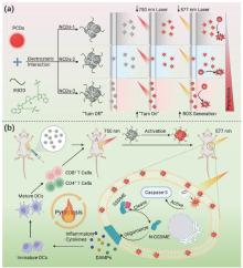
Scheme1
Schematic diagrams illustrating (a) the preparation of NCDs, the PDT regimen, (b) the induction of pyroptosis, and the antitumor immune response."
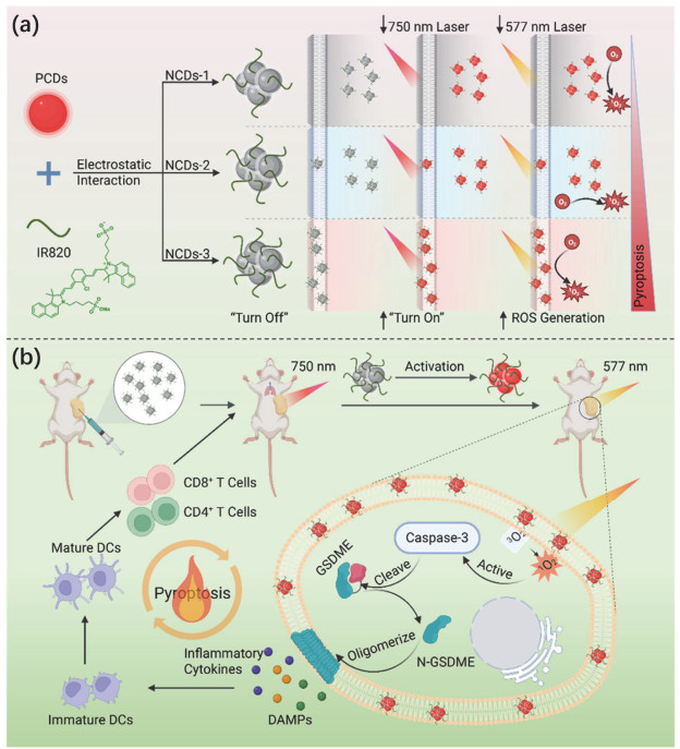
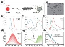
Fig 1
Characterization of NCDs. (a) The preparation diagram of NCDs. (b) The TEM image of NCDs. Scale bar: 200 nm. (c) The DLS analysis of PCDs and NCDs. (d) The absorption spectra of PCDs, IR820, and NCDs. (e) The NIR fluorescence spectra of IR820 and NCDs. (f) The fluorescence spectra of NCDs under 750 nm laser irradiation at different time (0–5 min). (g) The absorption spectra of NCDs under 750 nm laser irradiation at different time (0–5 min). (h) The 1O2 production of NCDs under different laser irradiation detected by ABDA indicator."
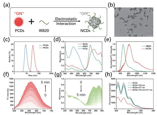
Fig 2
Cell membrane targeting property of NCDs. (a) The CLSM images of different NCDs incubated tumor cells with DiO. Green channel: 500–530 nm, excited at 488 nm; Red channel: 570–620 nm, excited at 561 nm. Scale bar: 50 μm. (b) The Pearson's coefficient between different NCDs and DiO. (c) The zeta potential of different NCDs. (d) The contact angle of different NCDs with H2O. (e) The images of contact angle with H2O."
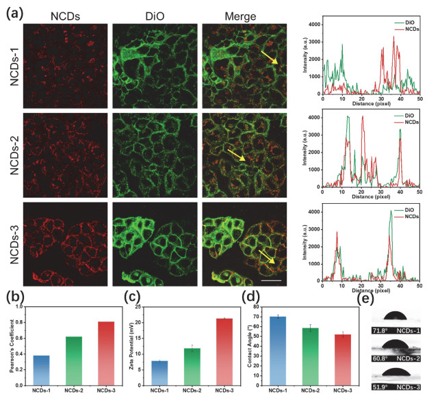
Fig 3
Cell pyroptosis induced by the membrane targeting NCDs. (a) Cell viability of tumor cells after PDT with different NCDs. (b) The LDH release from cell supernatant after PDT with different NCDs. (c) The ATP release from cell supernatant after PDT with different NCDs. (d) Western blotting analysis of pyroptosis related proteins expression in cells. (e) The ratios of C-GSDME to β-actin. (f) The ratios of C-Caspase3 to β-actin. (g) Confocal images of PI (red) and Annexin V-FITC (green) staining after different NCDs treatments. Green channel: 500–530 nm, excited at 488 nm; Red channel: 570–620 nm, excited at 561 nm. Scale bar: 50 μm."
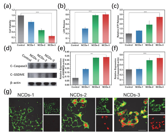
Fig 4
Photo-controlled pyroptosis and its immunogenicity. (a) Cell viability of tumor cells after different treatments. (b) Representative phase-contrast cell images after different treatments. Scale bar: 50 μm. (c) The LDH release from cell supernatant after different treatments. (d) Western blotting analysis of pyroptosis related proteins expression in cells after different treatments. (e) The ratios of C-GSDME to β-actin. (f) The ratios of C-Caspase3 to β-actin. (g) Confocal images of PI (red) and Annexin V-FITC (green) staining after NCDs + 750 nm laser + 577 nm laser. Green channel: 500–530 nm, excited at 488 nm; Red channel: 570–620 nm, excited at 561 nm. Scale bar: 50 μm. (h) Quantitative percentage of PI and Annexin V-FITC positive cells by flow cytometry. (i) Representative CLSM images of HMGB1 on tumor cells after different treatments. Blue channel: 433–468 nm, excited at 405 nm; Green channel: 500–530 nm, excited at 488 nm. Scale bar: 50 μm. (j) Quantification mean fluorescence intensity of HMGB1 in tumor cells. (k) Quantification mean fluorescence intensity of CRT in tumor cells. (l) The relative amounts of released ATP after different treatments. Ⅰ: Control, Ⅱ: NCDs, Ⅲ: NCDs + 750 nm laser, 100 mW cm−2, Ⅳ: NCDs + 577 nm laser, 50 mW cm−2, and Ⅴ: NCDs + 750 nm laser, 100 mW cm−2 + 577 nm laser, 50 mW cm−2."
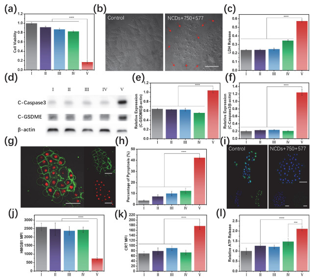
Fig 5
Antitumor effect of NCDs in vivo. (a) The NIR-Ⅱ fluorescence imaging of mice before and after NCDs injection. (b) The quantification of mean fluorescence intensity from A. (c) The fluorescence images of mice with NCDs injection before and after 750 nm laser irradiation. (d) The quantification of mean fluorescence intensity from C. (e) Schematic illustration showing the establishment of the 4T1 model for in vivo anti-tumor studies. (f) Photograph of tumors excised from mice with different treatments. (g) Tumor volume growth curves of mice with different treatments. (h) Tumor weight of mice in different groups. (i) Body weight curves of mice with different treatments. (j) H&E staining of tumor slices after different treatments. Scale bar: 100 μm. (k) H&E staining of lung metastatic nodules. Scale bar: 200 μm. Ⅰ: Control, Ⅱ: NCDs, Ⅲ: NCDs + 750 nm laser, 100 mW cm−2, Ⅳ: NCDs + 577 nm laser, 50 mW cm−2, and Ⅴ: NCDs + 750 nm laser, 100 mW cm−2 + 577 nm laser, 50 mW cm−2."
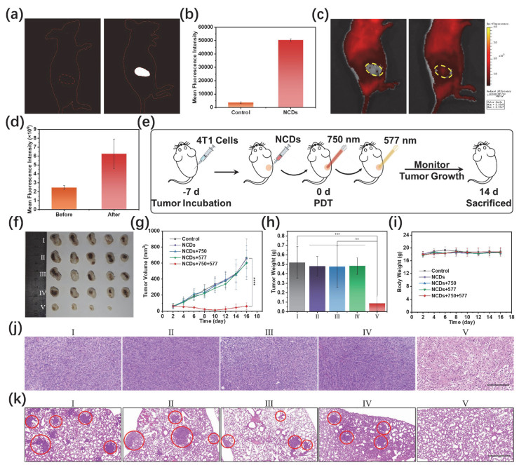
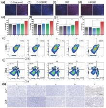
Fig 6
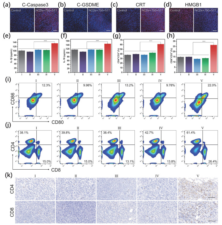
The immune response in vivo. (a) Immunofluorescence staining of C-Caspase3 protein in tumors after different treatments. Scale bar: 100 μm. (b) Immunofluorescence staining of C-GSDEM protein in tumors after different treatments. Scale bar: 100 μm. (c) Immunofluorescence analysis of CRT expression in tumors after different treatments. Scale bar: 100 μm. (d) Immunofluorescence analysis of HMGB1 expression in tumors after different treatments. Scale bar: 100 μm. (e) The levels of IL-1β in serum after different treatments. (f) The levels of IL-18 in serum after different treatments. (g) Representative flow cytometry of mature DCs in the spleen of mice. (h) Representative quantification of CD4+ T cells in the spleens of mice. (i) Representative quantification of CD8+ T cells in the spleens of mice. (j) Representative flow cytometry of CD8+/CD4+ T cells in the spleen of mice. (k) IHC analysis of tumor infiltrating CD8+/CD4+ T cells. Scale bar: 500 μm. Ⅰ: Control, Ⅱ: NCDs, Ⅲ: NCDs + 750 nm laser, 100 mW cm−2, Ⅳ: NCDs + 577 nm laser, 50 mW cm−2, and Ⅴ: NCDs + 750 nm laser, 100 mW cm−2 + 577 nm laser, 50 mW cm−2."
| 1 |
Y. Wang, K. Ma, M. Kang, D. Yan, N. Niu, S. Yan, P. Sun, L. Zhang, L. Sun, D. Wang, et al.. Chem. Soc. Rev. 2024, 53, 12014.
doi: 10.1039/D4CS00708E |
| 2 |
E. Nestoros, A. Sharma, E. Kim, J. S. Kim, M. Vendrell. Nat. Rev. Chem. 2025, 9, 46.
doi: 10.1038/s41570-024-00662-7 |
| 3 |
T. C. Pham, V. N. Nguyen, Y. Choi, S. Lee, J. Yoon. Chem. Rev. 2021, 121, 13454.
doi: 10.1021/acs.chemrev.1c00381 |
| 4 |
J. Kang, H. Jeong, M. Jeong, J. Kim, S. Park, J. Jung, J. M. An, D. Kim. J. Am. Chem. Soc. 2023, 145, 27587.
doi: 10.1021/jacs.3c09339 |
| 5 |
L. Li, X. Zhang, Y. Ren, Q. Yuan, Y. Wang, B. Bao, M. Li, Y. Tang. J. Am. Chem. Soc. 2024, 146, 5927.
doi: 10.1021/jacs.3c12132 |
| 6 |
X. Qu, F. Yin, M. Pei, Q. Chen, Y. Zhang, S. Lu, X. Zhang, Z. Liu, X. Li, H. Chen, et al.. ACS Nano 2023, 17, 11466.
doi: 10.1021/acsnano.3c01308 |
| 7 |
L. Feng, D. Tao, Z. Dong, Q. Chen, Y. Chao, Z. Liu, M. Chen. Biomaterials 2017, 127, 13.
doi: 10.1016/j.biomaterials.2016.11.027 |
| 8 |
X. Zhang, J. Xiong, K. Wang, H. Yu, B. Sun, H. Ye, Z. Zhao, N. Wang, Y. Wang, S. Zhang, et al.. Bioact. Mater. 2021, 6, 2291.
doi: 10.1016/j.bioactmat.2021.01.004 |
| 9 |
M. Dirak, C. M. Yenici, S. Kolemen. Coord. Chem. Rev. 2024, 506, 215710.
doi: 10.1016/j.ccr.2024.215710 |
| 10 |
J. B. Zhuang, Z. D. Ma, N. Li, H. Chen, L. J. Yang, Y. Lu, K. Y. Guo, N. Zhao, B. Z. Tang. Adv. Mater. 2024, 36, 2309488.
doi: 10.1002/adma.202309488 |
| 11 |
F. H. Igney, P. H. Krammer. Nat. Rev. Cancer 2002, 2, 277.
doi: 10.1038/nrc776 |
| 12 |
K. Hadian, B. R. Stockwell. Nat. Rev. Drug Discov. 2023, 22, 723.
doi: 10.1038/s41573-023-00749-8 |
| 13 |
X. Sun, M. Li, P. Wang, Q. Bai, X. Cao, D. Mao. Small Methods 2023, 7, 2201614.
doi: 10.1002/smtd.202201614 |
| 14 |
W. T. Gao, X. Y. Wang, Y. Zhou, X. Q. Wang, Y. Yu. Sig. Transdut. Target. Ther. 2022, 7, 196.
doi: 10.1038/s41392-022-01046-3 |
| 15 |
P. Fontana, G. Du, Y. Zhang, H. Zhang, S. M. Vora, J. J. Hu, M. Shi, A. B. Tufan, L. B. Healy, S. Xia, et al.. Cell 2024, 187, 6165.
doi: 10.1016/j.cell.2024.08.007 |
| 16 |
Q. Y. Wang, Y. P. Wang, J. J. Ding, C. H. Wang, X. H. Zhou, W. Q. Gao, H. W. Huang, F. Shao, Z. B. Liu. Nature 2020, 579, 421.
doi: 10.1038/s41586-020-2079-1 |
| 17 |
Z. B. Zhang, Y. Zhang, S. Y. Xia, Q. Kong, S. Y. Li, X. Liu, C. Junqueira, K. F. Meza-Sosa, T. M. Y. Mok, J. Ansara, et al.. Nature 2020, 579, 415.
doi: 10.1038/s41586-020-2071-9 |
| 18 |
Z. He, D. Feng, C. Zhang, Z. Chen, H. Wang, J. Hou, S. Li, X. Wei. J. Control. Release 2024, 366, 375.
doi: 10.1016/j.jconrel.2023.12.023 |
| 19 |
M. Wu, X. G. Liu, H. Chen, Y. K. Duan, J. J. Liu, Y. T. Pan, B. Liu. Angew. Chem. Int. Ed. 2021, 60, 9093.
doi: 10.1002/anie.202016399 |
| 20 |
M. Li, J. Kim, H. Rha, S. Son, M. S. Levine, Y. Xu, J. L. Sessler, J. S. Kim. J. Am. Chem. Soc. 2023, 145, 6007.
doi: 10.1021/jacs.3c01231 |
| 21 |
M. Wang, M. Wu, X. Liu, S. Shao, J. Huang, B. Liu, T. Liang. Adv. Sci. 2022, 9, 2202914.
doi: 10.1002/advs.202202914 |
| 22 |
Z. G. Yi, X. J. Qin, L. Zhang, H. Chen, T. L. Song, Z. C. Luo, T. Wang, J. Lau, Y. L. Wu, T. B. Toh, et al.. J. Am. Chem. Soc. 2024, 146, 9413.
doi: 10.1021/jacs.4c01929 |
| 23 |
Q. Liu, X. Yao, L. Zhou, W. Wu, J. Cheng, Z. Zhang, Z. Li, H. Sun, J. Jin, M. Zhang, et al.. Adv. Mater. 2024, 36, 2401145.
doi: 10.1002/adma.202401145 |
| 24 |
X. Wu, J. J. Hu, J. Yoon. Angew. Chem. Int. Ed. 2024, 63, e202400249.
doi: 10.1002/anie.202400249 |
| 25 |
C. Xiang, Y. Liu, Q. Ding, T. Jiang, C. Li, J. Xiang, X. Yang, T. Yang, Y. Wang, Y. Tan, et al.. Adv. Funct. Mater. 2024, 35, 2417979.
doi: 10.1002/adfm.202417979 |
| 26 |
Z. Li, F. Mo, Y. Wang, W. Li, Y. Chen, J. Liu, T.-J. Chen-Mayfield, Q. Hu. Nat. Commun. 2022, 13, 6321.
doi: 10.1038/s41467-022-34036-8 |
| 27 |
Y. Q. Tang, H. K. Bisoyi, X. M. Chen, Z. Y. Liu, X. Chen, S. Zhang, Q. Li. Adv. Mater. 2023, 35, 2300232.
doi: 10.1002/adma.202300232 |
| 28 |
Y. Liu, Z. Huang, X. Wang, Y. Hao, J. Yang, H. Wang, S. Qu. Adv. Funct. Mater. 2025, 35, 2420587.
doi: 10.1002/adfm.202420587 |
| 29 |
B. Y. Wang, S. Y. Lu. Matter 2022, 5, 110.
doi: 10.1016/j.matt.2021.10.016 |
| 30 |
L. Jiang, H. Cai, W. W. Zhou, Z. J. Li, L. Zhang, H. Bi. Adv. Mater. 2023, 35, 2210776.
doi: 10.1002/adma.202210776 |
| 31 |
J. C. Ge, M. H. Lan, B. J. Zhou, W. M. Liu, L. Guo, H. Wang, Q. Y. Jia, G. L. Niu, X. Huang, H. Y. Zhou, et al.. Nat. Commun. 2014, 5, 4596.
doi: 10.1038/ncomms5596 |
| 32 |
S. H. Li, W. Su, H. Wu, T. Yuan, C. Yuan, J. Liu, G. Deng, X. C. Gao, Z. M. Chen, Y. M. Bao, et al.. Nat. Biomed. Eng. 2020, 4, 704.
doi: 10.1038/s41551-020-0540-y |
| 33 |
L. Ðorđević F. Arcudi, M. Cacioppo, M. Prato. Nat. Nanotechnol. 2022, 17, 112.
doi: 10.1038/s41565-021-01051-7 |
| 34 |
T. Han, Y. Wang, S. Ma, M. Li, N. Zhu, S. Tao, J. Xu, B. Sun, Y. Jia, Y. Zhang, et al.. Adv. Sci. 2022, 9, 2203474.
doi: 10.1002/advs.202203474 |
| 35 |
H. Sun, S. Sun, H. Wang, K. Cheng, Y. Zhou, X. Wang, S. Gao, J. Mo, S. Li, H. Lin, et al.. Acta Biomater. 2025, 194, 352.
doi: 10.1016/j.actbio.2025.01.030 |
| 36 |
Y. Zhang, Q. Jia, F. Nan, J. Wang, K. Liang, J. Li, X. Xue, H. Ren, W. Liu, J. Ge, et al.. Biomaterials 2023, 293, 121953.
doi: 10.1016/j.biomaterials.2022.121953 |
| 37 |
X. Zhang, L. Li, B. Wang, Z. Cai, B. Zhang, F. Chen, G. Xing, K. Li, S. Qu. Angew. Chem. Int. Ed. 2024, 63, e202410522.
doi: 10.1002/anie.202410522 |
| 38 |
B. Wang, G. I. N. Waterhouse, B. Yang, S. Lu. Acc. Chem. Res. 2024, 57, 2928.
doi: 10.1021/acs.accounts.4c00516 |
| 39 |
Q. Cheng, T. Zhang, Q. Wang, X. Wu, L. Li, R. Lin, Y. Zhou, S. Qu. Adv. Mater. 2024, 36, 2408685.
doi: 10.1002/adma.202408685 |
| 40 |
T. Chen, K. Liang, J. Wang, J. Li, X. Xue, Y. Hao, H. Liang, H. Ren, H. Xiao, J. Ge, et al.. Nano Lett. 2024, 24, 14709.
doi: 10.1021/acs.nanolett.4c03913 |
| 41 |
J. Yang, B. Ren, X. Yin, L. Xiang, Y. Hua, X. Huang, H. Wang, Z. Mao, W. Chen, J. Deng. Adv. Mater. 2024, 36, 2402720.
doi: 10.1002/adma.202402720 |
| 42 |
Y. J. Li, Y. C. Guo, K. X. Zhang, R. R. Zhu, X. Y. Chen, Z. Z. Zhang, W. J. Yang. Adv. Sci. 2024, 11, 2306580.
doi: 10.1002/advs.202306580 |
| 43 |
J. Xu, X. Zheng, T.-B. Ren, L. Shi, X. Yin, L. Yuan, X.-B. Zhang. Coord. Chem. Rev. 2025, 528, 216379.
doi: 10.1016/j.ccr.2024.216379 |
| 44 |
Y. Li, D. Hu, M. Pan, Y. Qu, B. Chu, J. Liao, X. Zhou, Q. Liu, S. Cheng, Y. Chen, et al.. Biomaterials 2022, 288, 121700.
doi: 10.1016/j.biomaterials.2022.121700 |
| 45 |
D.-T. Nguyen, M.-J. Baek, S. M. Lee, D. Kim, S.-Y. Yoo, J.-Y. Lee, D.-D. Kim. Nat. Nanotechnol. 2024, 19, 1723.
doi: 10.1038/s41565-024-01757-4 |
| 46 |
M. Yu, S. Li, X. Ren, N. Liu, W. Guo, J. Xue, L. Tan, C. Fu, Q. Wu, M. Niu, et al.. ACS Nano 2024, 18, 3636.
doi: 10.1021/acsnano.3c11433 |
| 47 |
H. Xia, W. Zhou, D. Li, F. Peng, L. Yu, Y. Sang, H. Liu, A. Hao, J. Qiu. Angew. Chem. Int. Ed. 2024, 63, e202312755.
doi: 10.1002/anie.202312755 |
| 48 |
T. Zhang, X. Yang, X. Ou, M. M. S. Lee, J. Zhang, C. Xu, X. Yu, P. Gong, J. W. Y. Lam, P. Zhang, et al.. Adv. Mater. 2023, 35, 2303186.
doi: 10.1002/adma.202303186 |
| 49 |
L. H. Liu, W. X. Qiu, Y. H. Zhang, B. Li, C. Zhang, F. Gao, L. Zhang, X. Z. Zhang. Adv. Funct. Mater. 2017, 27, 1700220.
doi: 10.1002/adfm.201700220 |
| 50 |
S. J. Hao, Y. X. Zhu, F. G. Wu. J. Control. Release. 2023, 357, 222.
doi: 10.1016/j.jconrel.2023.03.038 |
| 51 |
K. Minton. Nat. Rev. Mmunol. 2020, 20, 274.
doi: 10.1038/s41577-020-0297-2 |
| [1] | Renyi Shao, Khurram Abbas, Vladimir Yu. Osipov, Haimei Zhu, Yuan Li, Usama, Hong Bi. Red-emitting carbon dots prepared from Epipremnum Aureum leaves extract for biological imaging [J]. Acta Phys. -Chim. Sin., 2026, 42(2): 100134-. |
| [2] | Yu Liu, Pengfei Li, Yize Liu, Zaicheng Sun. Recent advances in carbon dots as a single photocatalyst [J]. Acta Phys. -Chim. Sin., 2026, 42(2): 100167-. |
| [3] | Zihan Cheng, Kai Jiang, Jun Jiang, Henggang Wang, Hengwei Lin. Achieving thermal-stimulus-responsive dynamic afterglow from carbon dots by singlet-triplet energy gap engineering through covalent fixation [J]. Acta Phys. -Chim. Sin., 2026, 42(2): 100169-. |
| [4] | Chunyuan Kang, Xiaoyu Li, Fan Yang, Bai Yang. Ionic-bond crosslinked carbonized polymer dots for tunable and enhanced room temperature phosphorescence [J]. Acta Phys. -Chim. Sin., 2026, 42(1): 100156-. |
| [5] | Xue Wu, Yupeng Liu, Bingzhe Wang, Lingyun Li, Zhenjian Li, Qingcheng Wang, Quansheng Cheng, Guichuan Xing, Songnan Qu. Rationally assembling different surface functionalized carbon dots for enhanced near-infrared tumor photothermal therapy [J]. Acta Phys. -Chim. Sin., 2025, 41(9): 100109-. |
| [6] | Wenlong Wang, Wentao Hao, Lang He, Jia Qiao, Ning Li, Chaoqiu Chen, Yong Qin. Bandgap and adsorption engineering of carbon dots/TiO2 S-scheme heterojunctions for enhanced photocatalytic CO2 methanation [J]. Acta Phys. -Chim. Sin., 2025, 41(9): 100116-. |
| [7] | Jianqiao Chang, Huimin Xu, Wenjing Xie, Yang Zhang, Ling Qi, Louzhen Fan, Yong Li. Fluorescent Carbon Dots for Rapid and Highly Sensitive Detection of Nucleic Acids [J]. Acta Phys. -Chim. Sin., 2023, 39(12): 2301034-. |
| [8] | Yuan Liu, Weidong Li, Han Wu, Siyu Lu. Carbon Dots Enhance Ruthenium Nanoparticles for Efficient Hydrogen Production in Alkaline [J]. Acta Phys. -Chim. Sin., 2021, 37(7): 2009082-. |
| [9] | Yanjie HUANG, Chao LIAN, Jinyan ZHOU, Zichen HUANG, Xiaohong KANG, Zhenyu HUANG, Xiaojing LI, Lin CHEN, Yan GUAN. Investigation of Excitation-, pH-, Metal Ion-, Temperature-, and Polarity-Dependent Fluorescence of Carbon Dots Derived from Silkworm Excrement [J]. Acta Physico-Chimica Sinica, 2019, 35(11): 1267-1275. |
| [10] | Jin-Long LIU,Liang-Zhen LIN,Jin-Feng HU,Ming-Jie BAI,Liang-Xian CHEN,Jun-Jun WEI,Li-Fu HEI,Cheng-Ming LI. Reaction Process and Luminescence Mechanism of Carbon Nanodots Prepared by Microwave Synthesis [J]. Acta Phys. -Chim. Sin., 2018, 34(1): 92-98. |
| [11] | Li-Xia SANG,Jia LIN,Hao GE,Lei LEI. Dynamic Analysis of Carbon Dots/KOH Electrolyte Interface by IMPS/IMVS [J]. Acta Phys. -Chim. Sin., 2017, 33(12): 2454-2462. |
|
||